. 2009;189(1-4):144-52.
doi: 10.1159/000151747. Epub 2008 Aug 27.

Transcriptional regulation of osteoblasts

Affiliations

Transcriptional regulation of osteoblasts

Renny T Franceschi et al. Cells Tissues Organs. 2009.

Abstract

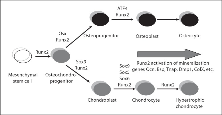
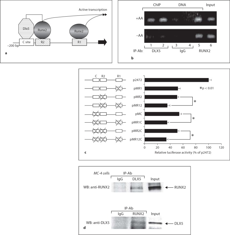
The differentiation of osteoblasts from mesenchymal precursors requires a series of cell fate decisions controlled by a hierarchy of transcription factors. These include RUNX2, Osterix (OSX), ATF4 and a large number of nuclear coregulators. During bone development, initial RUNX2 expression coincides with the formation of mesenchymal condensations and precedes the branching of chondrogenic and osteogenic lineages. Given its central role in bone development, it is not surprising that RUNX2 is subject to a variety of controls. These include posttranslational modification, especially phosphorylation, and interactions with accessory nuclear factors. Specific examples of RUNX2 regulation to be reviewed include phosphorylation by the ERK/MAP kinase pathway and interactions with DLX5. RUNX2 is regulated via phosphorylation of critical serine residues in the proline/serine/threonine domain. In vivo, the transgenic expression of constitutively active MAP kinase in osteoblasts accelerated skeletal development, while a dominant-negative MAPK retarded development in a RUNX2-dependent manner. DLX5-RUNX2 complexes can be detected in osteoblasts and this interaction plays a critical role in maintaining osteoblast-specific expression of the bone sialoprotein gene. These studies allow us to begin understanding the complex mechanisms necessary to fine-tune bone formation as mesenchymal progenitors progress down the osteoblast lineage.


Figures

Fig. 1
Fig. 1
Transcription factor control of skeletal lineages. Major transcription factors that, based on genetic studies, are involved in osteoblast and chondrocyte differentiation are included in this chart. Also shown is the sequential nature of transcription factor expression with RUNX2 persisting throughout osteoblast and chondrocyte lineages [Franceschi et al., 2007].
Fig. 2
Fig. 2
RUNX2 protein levels are not well correlated with transcriptional activity. MC3T3-E1 clone 4 preosteoblast cells were grown in control (–) or ascorbate-containing medium (+). At the times indicated, RUNX2 protein levels were determined by Western blotting (a), while Runx2 (Osf2), Ocn and Bsp mRNA levels (b–d) were measured on Northern blots (b) and quantified by densitometry. c Runx2. d Ocn and Bsp. Open symbols = Control; closed symbols = ascorbate. ○, • = Ocn mRNA; •, █ = Bsp mRNA [Xiao et al. 1998].
Fig. 3
Fig. 3
Altered skeletal development in TgMek-dn and TgMek-sp mice. a Whole mounts of E15.5 skeletons stained with alcian blue and alizarin red. d Effects of transgene expression on embryo weights. b, e Cranial bones showing differences in mineralization (b) and quantification of mineralized area (expressed as percent of total calvarial area) (e). c, f Hindlimbs showing differences in the size of bones with transgene expression (c) and quantification of femur lengths (f). g Histology of long bones from wild-type, TgMek-dn and TgMek-sp mice. Note the delay in bony collar and trabecular bone in TgMek-dn embryos. Statistical analysis: values are expressed as means ± SD, n = 8/group. * Significantly different from wild type at p < 0.01. Reproduced from Ge et al. [2007].
Fig. 4
Fig. 4
Genetic interactions between Mek-dn and Mek-sp transgenes and Runx2.TgMek-dn or TgMek-sp mice were crossed with RUNX2+/– mice to generate the genotypes indicated. a–d Partial rescue of cleidocranial dysplasia phenotype in RUNX2+/– mice with Mek-sp. a Skeletal whole mounts of newborn mice stained with alcian blue and alizarin red (top), isolated clavicles (middle) and crania (bottom). b–d Measurements of femur length (b), clavicle areas (c) and mineralized area of calvaria (expressed as a fraction of total calvarial area) (d). e–h Increased severity of cleidocranial dysplasia phenotype with Mek-dn. Groups are as in panels a–d. Statistical analysis: values are expressed as means ± SD, n = 8/group. Comparisons are indicated by bars. * p < 0.01. Reproduced from Ge et al. [2007].
Fig. 5
Fig. 5
In vivo binding of RUNX2 and DLX5 to chromatin sites in differentiated and undifferentiated cells. a Schematic of the proximal Bsp promoter. RUNX2 and homeodomain (DLX5) protein-binding sites are indicated. b Comparison of Bsp chromatin occupancy by RUNX2 and DLX5. Chromatin immunoprecipitation assays were used to detect RUNX2 and DLX5 bound to the proximal Bsp promoter in control (–AA) and differentiated (+AA) MC3T3-E1 clone 4 cells. Antibodies used for chromatin immunoprecipitation are indicated. Note that RUNX2 remains chromatin associated regardless of differentiated state, while Dlx5 is only present in differentiated cells. c Functional interaction between R2 and C sites. 2.5-kb Bsp-luc reporter constructs containing all possible combinations of R1, R2 and C site mutations were transfected into MC3T3-E1 cl4 cells and grown under differentiating conditions. Note that mutation of either R2 or C is as inhibitory as mutation of both sites, while mutation of either site together with R1 gives maximal inhibition. d Protein-protein interactions between DLX5 and RUNX2. Pull-down assays with nuclear extracts from differentiated MC3T3 cells were used to show physical association between these 2 factors [Roca et al., 2005]. ChIP = Chromatin immunoprecipitation; IP-Ab = chromatin immunoprecipitation antibodies; WB = Western blot.

References

    1. Afzal F., Pratap J., Ito K., Ito Y., Stein J.L., van Wijnen A.J., Stein G.S., Lian J.B., Javed A. Smad function and intranuclear targeting share a Runx2 motif required for osteogenic lineage induction and BMP2 responsive transcription. J Cell Physiol. 2005;204:204–63. - PubMed
    1. Benson M.D., Bargeon J.L., Xiao G., Thomas P.E., Kim A., Cui Y., Franceschi R.T. Identification of a homeodomain binding element in the bone sialoprotein gene promoter that is required for its osteoblast-selective expression. J Biol Chem. 2000;275:275–13907. - PubMed
    1. Bialek P., Kern B., Yang X., Schrock M., Sosic D., Hong N., Wu H., Yu K., Ornitz D.M., Olson E.N., Justice M.J., Karsenty G. A twist code determines the onset of osteoblast differentiation. Dev Cell. 2004;6:6–423. - PubMed
    1. Ducy P., Zhang R., Geoffroy V., Ridall A.L., Karsenty G. Osf2/Cbfa1 a transcriptional activator of osteoblast differentiation. Cell. 1997;89:89–747. - PubMed
    1. Engler A.J., Sen S., Sweeney H.L., Discher D.E. Matrix elasticity directs stem cell lineage specification. Cell. 2006;126:126–677. - PubMed

MeSH terms