Review
. 2008 May 28;6(2):180-219.
doi: 10.3390/md20080010.

Domoic acid toxicologic pathology: a review

Affiliations
Review

Domoic acid toxicologic pathology: a review

Olga M Pulido. Mar Drugs. .

Abstract

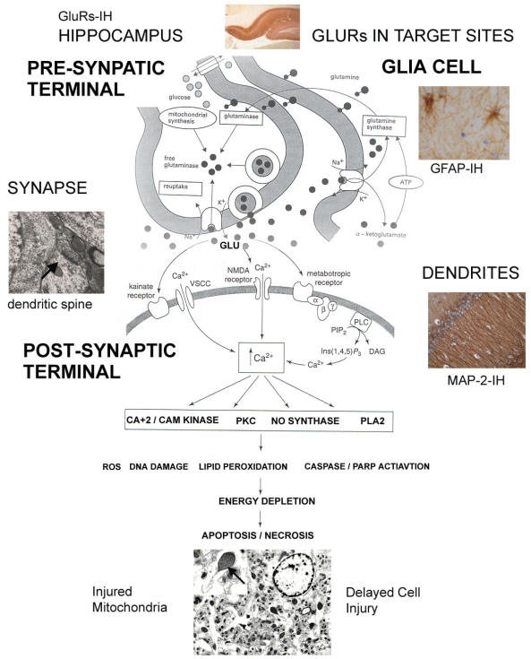
Domoic acid was identified as the toxin responsible for an outbreak of human poisoning that occurred in Canada in 1987 following consumption of contaminated blue mussels [Mytilus edulis]. The poisoning was characterized by a constellation of clinical symptoms and signs. Among the most prominent features described was memory impairment which led to the name Amnesic Shellfish Poisoning [ASP]. Domoic acid is produced by certain marine organisms, such as the red alga Chondria armata and planktonic diatom of the genus Pseudo-nitzschia. Since 1987, monitoring programs have been successful in preventing other human incidents of ASP. However, there are documented cases of domoic acid intoxication in wild animals and outbreaks of coastal water contamination in many regions world-wide. Hence domoic acid continues to pose a global risk to the health and safety of humans and wildlife. Several mechanisms have been implicated as mediators for the effects of domoic acid. Of particular importance is the role played by glutamate receptors as mediators of excitatory neurotransmission and the demonstration of a wide distribution of these receptors outside the central nervous system, prompting the attention to other tissues as potential target sites. The aim of this document is to provide a comprehensive review of ASP, DOM induced pathology including ultrastructural changes associated to subchronic oral exposure, and discussion of key proposed mechanisms of cell/tissue injury involved in DOM induced brain pathology and considerations relevant to food safety and human health.

Keywords: Amnesic Shellfish Poisoning; Domoic Acid; Excitotoxicity; Food Safety; Glutamate Receptors; Neuropathology; Neurotoxicology; Toxicologic Pathology.


Figures

Figure 1
Figure 1
Brain of a control rat after trans-cardiac perfusion with heparinized Tyrode=s solution followed by 10% neutral buffered formalin. All images are from paraffin sections stained with H&E. A) Cross section of a hippocampus showing the granular cell layer (GL) of the dentate gyrus (DG), the molecular layer (ML), the CA3 and the CA4 regions. Objective x10. B) Shows the CA3 region with well preserved pyramidal cells (Py). Blood vessels are seen as white spaces with the endothelial cells at the periphery (arrow). Objective x40. C) Cross section of both hippocampal formations (Hi) showing the dentate gyrus (DG), the CA3, CA2 and CA1 regions. This image is a scanned mid coronal section of the rat brain. For anatomical reference see technical notes.
Figure 2
Figure 2
Brain of a rat after trans-cardiac perfusion with heparinized Tyrode=s solution followed by 10% neutral buffered formalin. A) Sections of the hippocampus of a rat treated with 4 mg/kg bw/ip of DOM showing cell drop out and neuronal necrosis, particularly within the CA3 regions. H&E. Objective x5. B). Higher magnification of the CA3 region showing pyramidal neurons (Py) with vacuolar cytoplasm (V). Few shrunken neurons (*) and nuclear pyknosis (arrow) are also seen. H&E. Objective x40.
Figure 3
Figure 3
Brain of a rat after trans-cardiac perfusion with heparinized Tyrode=s solution followed by 10% neutral buffered formalin. A) Hippocampus of a control rat. CA3 regions. Chen/Bodian Objective x20. B) Sections of the hippocampus of a rat treated with 4mg/kg bw/ip of DOM, showing-marked drop out of pyramidal neurons in the CA3 region. Chen/Bodian, Objective x20.
Figure 4
Figure 4
Brain of monkeys (M. fascicularis) after trans-cardiac perfusion with heparinized Tyrode=s solution followed by 10% neutral buffered formalin. A) Section of the CA3 region of the hippocampus of a control animal showing well preserved pyramidal cells (Py). Blood vessels are seen as white spaces with the endothelial cells at the periphery (arrow), H&E Objective x40. B) Sections of the hippocampus of an animal treated with 4 mg/kg bw/ip of DOM showing cell drop out and neuronal necrosis. Most pyramidal neurons appear with vacuolar cytoplasm (V). Some nuclear pyknosis (arrow head) is also seen. H&E, Objective x40.
Figure 5
Figure 5
Brain of monkeys (M. fascicularis) after trans-cardiac perfusion with heparinized Tyrode=s solution followed by 10% neutral buffered formalin. Histological sections were processed for GFAP immunohistochemistry. A) Hippocampus of a control animal showing the granular cell layer (GL) of the dentate gyrus and the CA4 region showing spaced astrocytes labeled by the GFAP-IH. Some immunolabeled astrocytes are clearly identified around blood vessels (arrow), Objective x10. B) Hippocampus of an animal treated with a single IV dose (0.055 mg/kg/bw) of DOM. Animal recovered after the initial symptoms of toxicity which lasted 90 minutes and included vomiting, gagging, lethargy and disorientation. Necropsy was conducted six month after the injection. Sections show marked astrocytosis as revealed by the intensity of the GFAP-IH seen in the CA4 and subgranular zone (arrow); granular cell layer (GL). Objective x20
Figure 6
Figure 6
Retina of a monkey (M. fascicularis) fixed by intraocular injection of 10% neutral buffered formalin. Photographs show cross sections of the retina stained with H&E. Picture shows the retina of an animal treated with DOM (4mg/kg/bw ip). Cell loss and necrosis are present in the INL, GCL and to a lesser extent in the ONL. Vacuoles are easily identified in the Ph cell layer, particularly the cones (*) and in the OPL. Cones and rods in the Ph layer are identified (arrows). There is also marked loss of cell bodies in the GCL. From external to internal: the photoreceptor cell layer (Ph); the outer nuclear layer (ONL); the outer plexiform layer (OPL); inner nuclear layer (INL); inner plexiform layer (IPL); ganglion cell layer (GCL). Objective x40.
Figure 7
Figure 7
Brain of rats after trans-cardiac perfusion with heparinized Tyrode=s solution followed by a fixative containing 2% glutaraldehyde and 2% paraformaldehyde in Tyrode=s solution. Samples selected from the CA3 region were processed for electron microscopy (EM). A) CA3 region of the hippocampus of a control rat showing a cluster of pyramidal cells with good preservation and integrity of cell membranes and organelles. The nucleus (N) and nucleolus (ncl) are easily identified. A well preserved electro dense dendritic spine is depicted (arrow). Scale Bar = x2.5μm; B) CA3 region of the hippocampus of a rat treated by gavage with 5.0 mg/kg/day of DOM for 64 days [81]. Image shows a pyramidal cell with easily identifiable nucleus (N), the cytoplasm and surrounding neuropil with numerous vacuoles (V) of various sizes giving a >Swiss cheese’ effect. The neuropil refers to intricate interwoven cell processes including: glial processes, synaptic terminals, axons, and dendrites that are interspersed among the nerve cells in the gray matter of the CNS. Scale Bar = x2.5μm; C) CA3 region of the hippocampus of a control rat showing good preservation and integrity of the neuropil. Mitochondria (M), synaptic spines (arrow) and dendrites (D) are identified. Scale Bar = x1.1μm; D) CA3 region of the hippocampus of a rat treated by gavage with 5.0 mg/kg/day of DOM for 64 days. Vacuoles (V) and the ‘Swiss cheese’ effect are more apparent at this magnification. Some remaining dendritic spines (arrow) can still be identified. There is increased electron density of the mitochondria (M) with loss organization of the cristae. Scale Bar = x1.1μm; E) High magnification of the CA3 region of the hippocampus of a control rat showing good preservation and structural integrity. An electro dense dendritic spine (arrow), terminal axon (At), dendrite and mitochondria (M) are identified. Scale Bar = x 0.6μm; F). High magnification of the CA3 region of the hippocampus of a rat treated by gavage with 5.0 mg/kg/day of DOM for 64 days showing loss of structural integrity and marked vacuolar (V) dilatation of dendrites. Scale Bar = x 0.6μm
Figure 7
Figure 7
Brain of rats after trans-cardiac perfusion with heparinized Tyrode=s solution followed by a fixative containing 2% glutaraldehyde and 2% paraformaldehyde in Tyrode=s solution. Samples selected from the CA3 region were processed for electron microscopy (EM). A) CA3 region of the hippocampus of a control rat showing a cluster of pyramidal cells with good preservation and integrity of cell membranes and organelles. The nucleus (N) and nucleolus (ncl) are easily identified. A well preserved electro dense dendritic spine is depicted (arrow). Scale Bar = x2.5μm; B) CA3 region of the hippocampus of a rat treated by gavage with 5.0 mg/kg/day of DOM for 64 days [81]. Image shows a pyramidal cell with easily identifiable nucleus (N), the cytoplasm and surrounding neuropil with numerous vacuoles (V) of various sizes giving a >Swiss cheese’ effect. The neuropil refers to intricate interwoven cell processes including: glial processes, synaptic terminals, axons, and dendrites that are interspersed among the nerve cells in the gray matter of the CNS. Scale Bar = x2.5μm; C) CA3 region of the hippocampus of a control rat showing good preservation and integrity of the neuropil. Mitochondria (M), synaptic spines (arrow) and dendrites (D) are identified. Scale Bar = x1.1μm; D) CA3 region of the hippocampus of a rat treated by gavage with 5.0 mg/kg/day of DOM for 64 days. Vacuoles (V) and the ‘Swiss cheese’ effect are more apparent at this magnification. Some remaining dendritic spines (arrow) can still be identified. There is increased electron density of the mitochondria (M) with loss organization of the cristae. Scale Bar = x1.1μm; E) High magnification of the CA3 region of the hippocampus of a control rat showing good preservation and structural integrity. An electro dense dendritic spine (arrow), terminal axon (At), dendrite and mitochondria (M) are identified. Scale Bar = x 0.6μm; F). High magnification of the CA3 region of the hippocampus of a rat treated by gavage with 5.0 mg/kg/day of DOM for 64 days showing loss of structural integrity and marked vacuolar (V) dilatation of dendrites. Scale Bar = x 0.6μm
Figure 8
Figure 8
. Illustration of the key proposed mechanisms of tissue cell injury induced by DOM with photographs of target sites. The diagram shows the interaction between the presynaptic and the postsynaptic terminal. Glutamate receptors in target tissues such as the hippocampus are activated by EEAs such as DOM, which at the same time induces the release of glutamate. The figure shows vesicular release of glutamate into the synaptic space, activation of the post synaptic receptor systems, re-uptake into the pre synaptic terminal, and surrounding glia cells (astrocytes). DOM and glutamate activate the various glutamate receptors present in the postsynaptic membrane, inducing cellular injury through a common injury pathway, a process known as excitotoxicity. This process can be separated into several overlapping components:1)The iGluRs are ion-gated channels selective to Na+, K+ and Ca+2 and any sustained stimulation of these receptors may results in osmotic tissue damage. Hence, neuronal cell swelling reflects the influx of extra cellular Na+, Cl and water. Focal swellings along the dendrites called varicosities are early structural changes and are viewed as a hallmark of excitotoxic neuronal injury. 2) Activation of iGluRs triggers the influx of Ca+2 from the extra cellular environment to the synaptic cleft. This stage is marked by delayed cell degeneration. The accumulation of Ca+2 is the crucial determinant of injury. This elevation of Ca+2 triggers the activation of several enzymes: calmodulin (CAM), protein kinase (PKC), nitric oxide synthase (NO synthase), phospholipase A2 (PLA2) and reactive oxygen species (ROS). Evidence indicates that the mitochondrion plays a central role in the processes of excitotoxic neuronal cell degeneration with a web of interactions between Ca+2 homeostasis, ATP production, and the generation and detoxification of ROS. 3) GluRs are found localized at the synapse within electron dense structures known as postsynaptic density (PSD), mediating the binding of GluRs to sub membrane proteins such as actin and PDZ containing proteins. These proteins mediate protein-protein interactions. GluRs PDZ-containing proteins bind to numerous signal molecules including nitric oxide synthase, providing a mechanism for clustering GluRs with the corresponding signalling transduction protein. These GluRs associated proteins and excitotoxic signalling result in a free radical cascade and activation of enzymatic processes causing extensive damage of cell structures and ultimate cell death. 4) Astrocytes, as do neurons, express GluRs providing the binding effectors site for DOM and show degenerative structural changes in response to DOM exposure. Failure of astrocytes to remove extra cellular glutamate is one of the key compounding mechanisms implicated in DOM neurotoxicity. (Diagram - modified from Gill and Pulido [131])

References

    1. Hynie I, Hockin J, Wright J, Iverson F. Panel discussion: evidence that domoic acid was the cause of the 1987 outbreak. Can Dis Wkly Rep. 1990;16(Suppl 1E):37–40. - PubMed
    1. Todd EC. Chronology of the toxic mussels outbreak. Can Dis Wkly Rep. 1990;16(Suppl 1E):3–4. - PubMed
    1. Liston AJ. Domoic acid toxicity. Introduction. Can Dis Wkly Rep. 1990;16(Suppl 1E):1–2. - PubMed
    1. Perl TM, Bedard L, Kosatsky T, Hockin JC, Todd EC, Remis RS. An outbreak of toxic encephalopathy caused by eating mussels contaminated with domoic acid. N Engl J Med. 1990;322(25):1775–1780. - PubMed
    1. Quilliam MA, Wright JL. The amnesic shellfish poisoning mystery. Anal Chem. 1989;61(18):1053A–1106A. - PubMed

LinkOut - more resources