. 2008 Aug 27;3(8):e3077.
doi: 10.1371/journal.pone.0003077.

Drug-selected human lung cancer stem cells: cytokine network, tumorigenic and metastatic properties

Affiliations

Drug-selected human lung cancer stem cells: cytokine network, tumorigenic and metastatic properties

Vera Levina et al. PLoS One. .

Abstract

Background: Cancer stem cells (CSCs) are thought to be responsible for tumor regeneration after chemotherapy, although direct confirmation of this remains forthcoming. We therefore investigated whether drug treatment could enrich and maintain CSCs and whether the high tumorogenic and metastatic abilities of CSCs were based on their marked ability to produce growth and angiogenic factors and express their cognate receptors to stimulate tumor cell proliferation and stroma formation.

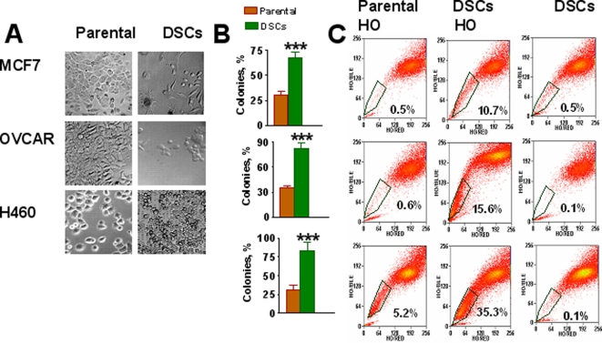
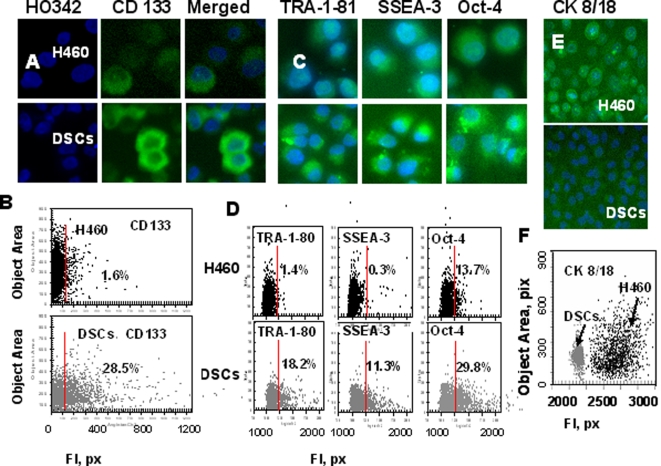
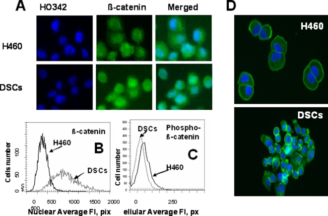
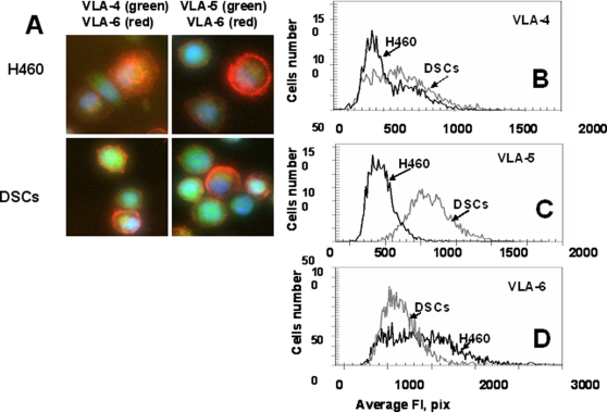
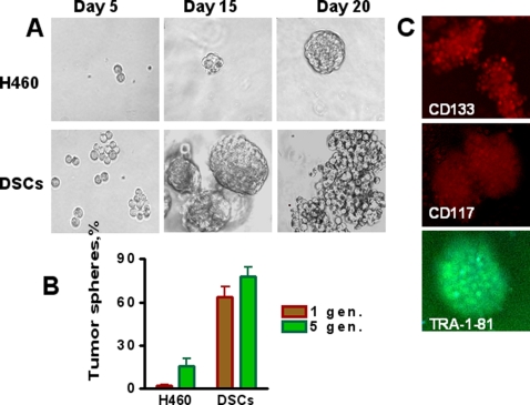
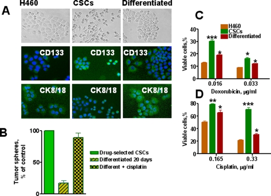
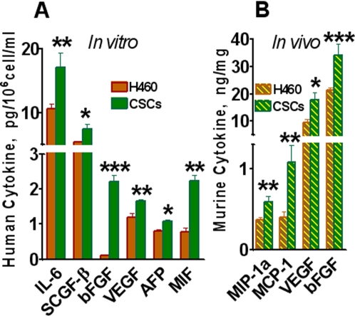
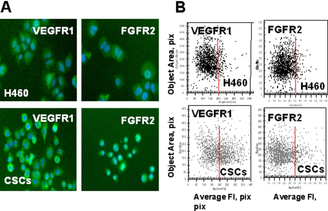
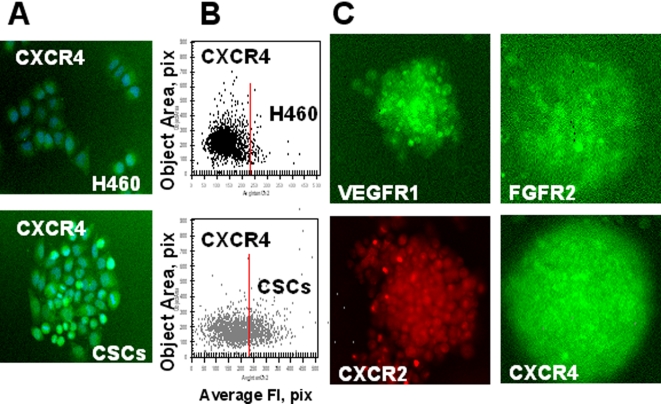
Methodology/findings: Treatment of lung tumor cells with doxorubicin, cisplatin, or etoposide resulted in the selection of drug surviving cells (DSCs). These cells expressed CD133, CD117, SSEA-3, TRA1-81, Oct-4, and nuclear beta-catenin and lost expression of the differentiation markers cytokeratins 8/18 (CK 8/18). DSCs were able to grow as tumor spheres, maintain self-renewal capacity, and differentiate. Differentiated progenitors lost expression of CD133, gained CK 8/18 and acquired drug sensitivity. In the presence of drugs, differentiation of DSCs was abrogated allowing propagation of cells with CSC-like characteristics. Lung DSCs demonstrated high tumorogenic and metastatic potential following inoculation into SCID mice, which supported their classification as CSCs. Luminex analysis of human and murine cytokines in sonicated lysates of parental- and CSC-derived tumors revealed that CSC-derived tumors contained two- to three-fold higher levels of human angiogenic and growth factors (VEGF, bFGF, IL-6, IL-8, HGF, PDGF-BB, G-CSF, and SCGF-beta). CSCs also showed elevated levels of expression of human VEGFR2, FGFR2, CXCR1, 2 and 4 receptors. Moreover, human CSCs growing in SCID mice stimulated murine stroma to produce elevated levels of angiogenic and growth factors.

Conclusions/significance: These findings suggest that chemotherapy can lead to propagation of CSCs and prevention of their differentiation. The high tumorigenic and metastatic potentials of CSCs are associated with efficient cytokine network production that may represent a target for increased efficacy of cancer therapy.


Conflict of interest statement

Competing Interests: The authors have declared that no competing interests exist.

Figures

Figure 1
Figure 1. Selection of DSCs populations from human carcinoma cell lines.
A, Morphology of parental MCF7, OVCAR3 and H460 cells and drug survived cells (DSCs). MCF-7 and H460 cells were treated with doxorubicin (0.125 µg/ml); OVCAR-3 cells were treated with cisplatin (3.3 µg/ml). After 48 h drugs were removed and drug-surviving cells (DSCs) were cultured for 3–4 weeks. B, Increased colony formation by DSCs isolated from parental MCF7 (breast), H460 (lung) and OVCAR-3 (ovarian) cancer cell lines. Cells were seeded 0.5 cell/per well in 96-well plates with culture media supplemented with 10% of FBS and cells were grown for two week. The percentage of colony formation was calculated. ***-P<0.001. C, Analysis of side population (SP) in DSCs and parental MCF7, OVCAR-3 and H460 cell lines. Tumor cells were stained with 5 µg/ml Hoechst33342 (HO). Some cells were pretreated with 10 µM fumitremorgin C (FTC) for 10 min prior to Hoechst addition (HO+FTC). Cells were resuspended in RPMI with 20% FBS and 2 µg/ml propidium iodide and sorted using MoFlo cytometer. Data for viable cells were analyzed for parametric correlations and annotated using FCS Express.
Figure 2
Figure 2. Analysis of CD133, embryonic stem cell (ESC) markers and cytokeratins 8/18 expression in H460 cells and DSCs.
H460 cells and DSCs, growing in 96-well plates, were fixed and incubated with primary Abs against CD133, TRA-1-81, SSEA-3, Oct-4, or cytokeratins8/18 and then with secondary Abs. Cell nuclei were stained with Hoechst 33342. Cell images were acquired using the Cellomics ArrayScan HCS Reader (20X, 40X objectives) and analyzed using the Target Activation BioApplication Software Module. A, Immunofluorescent images of tumor cells. B, Fluorescence intensity (pix) of CD133 plotted against object area. Each point represents a single cell. Cells to the right of the red line are CD133+ (above IgG control staining). C, Images of tumor cells immunofluorescently stained tumor cells for TRA-1-81, SSEA-3 and Oct-4 ES cell markers. D. Fluorescence intensity of TRA-1-81, SSEA-3 and Oct-4, plotted against object area. Each point represents a single cell. Cells to the right of the red line are positive (above IgG control staining). E,Images of immunofluorescently stained tumor cells for cytokeratins8/18. F, Fluorescence intensity of cytokeratins8/18 in H460 cells (black dots) and DSCs (grey dots) plotted against object area.
Figure 3
Figure 3. Analysis of β-catenin intracellular distribution in H460 cells and DSCs.
Cells were fixed and incubated with Alexa Fluor® 488 phalloidin or with primary Abs against β-catenin and with secondary Alexa Fluor 488 conjugated Abs. Next cells were stained with Hoechst33342. Cell images were acquired using the Cellomics ArrayScan HCS Reader (20X objective) and analyzed using the Compartment Analysis BioApplication Software Module and the Target Activation BioApplication Software Module. A, Images of H460 cells and DSCs immunofluorescently stained for β-catenin (A). B, An average fluorescence intensity of nuclear β-catenin in H460 (black line) and DSCs (grey line).C, An average fluorescence intensity of cellular phosphor- β-catenin in H460 (black line) and DSCs (grey line). D, Cytoskeleton images of H460 cells and DSCs immunofluorescently stained for phalloidin and Hoechst33342.
Figure 4
Figure 4. Expression of adhesion molecules, VLA-4(CD49d), VLA-5(CD49e), VLA-6(CD49f), by H460 cells and DSCs.
Cells were incubated with Abs against VLA-4-FITC and VLA-6-PC5 or VLA-5-FITC and VLA-6-PC5. Cell images were acquired using the Cellomics ArrayScan HCS Reader (20X, 40X objectives) and analyzed using the Target Activation BioApplication Software Module. A, Immunofluorescent images of VLA4/VLA6 (left) and VLA-5/VLA-6 (right) expression in H460 and DSCs cells (40X objective). B-D, An average fluorescence intensity of VLA-4(B), VLA-5(C) and VLA-6(D) in H460 cells (black line) and DSCs (grey line).
Figure 5
Figure 5. Chemotherapy selectively enriches for self-renewing lung cancer cells.
Parental H460 cells and DSCs (1000 cell/ml) were plated onto ultra low adherent plates in MC-based serum free media supplemented with growth factors and cultivated as described in Material and Methods. Tumor spheres generated from single-cell suspension cultures of parental H460 cells and drug survived CSCs were counted after 3 weeks of culture (1 st generation), and then spheres were dissociated and replated as described in Material and Methods. A, Lung tumor spheres generated from single-cell cultures of parental H460 cells and DSCs, imaged on indicated day of culture. B, Maintenance of enhanced ability to form tumor spheres during several generations of DSCs transfer (for comparison only 1-st and 5-th generation's data are presented). C, Immunofluorescent images of lung tumor spheres stained for CD133, CD117 and TRA-1-81 (10X objective).
Figure 6
Figure 6. In vitro differentiation potential of lung cancer sphere cells and drug resistance of CSCs.
A, Loss of stem cell marker (CD133) and increase of differentiation markers (CK8/18) by lung CSCs differentiated progenitors. Parental H460 cells and CSCs from tumor spheres were seeded in collagen coated well plates and cultured for 3 weeks in complete RPMI 1640 medium supplemented with 10% FBS. Upper row - cell images in phase –contrast microscopy; in the middle - cells immunofluorescently stained for CD133 and bottom row - cells immunofluorescently stained for CK 8/18. B, Self-renewing ability of differentiated lung cancer cells treated with cisplatin. Relative % of cells generated tumor spheres from single-cell suspension cultures of drug selected CSCs, cells differentiated during 3 weeks and Progenitors of CSCs differentiated for 3 weeks were treated with cisplatin (1 µM) for two days. Surviving cells were transferred into low adherent plates and cultured in semisolid serum free medium supplemented with growth factors. Numbers of formed tumor spheres were determined and presented as percent of control. Control is number of spheres formed by transfer of cells derived from control tumor spheres. Number of these spheres is accepted as 100 %. C, Effect of cisplatin and doxorubicin on proliferation of parental H460 cells, CSCs and their differentiated cells. H460, lung CSCs and differentiated cells were plated in 96-well plates precoated with Collagen at 1×10⁴ cells/well in complete RPMI 1640 medium with 10% FBS. After 24 h doxorubicin or cisplatin was added at the indicated concentrations. Cells were cultured for 72 h, fixed, stained with Hoechst 33342 (2 µg/mL), and counted using the Cellomics ArrayScan HCS Reader.
Figure 7
Figure 7. Multiplex analysis of cytokines.
A, In vitro cytokine production by CSCs and parental human tumor H460 cells. Cells were cultivated in 96-well plates for 24 h in complete RPMI 1640 medium; samples of conditioned media were collected. Cells were fixed, stained with Hoechst 33342, and cell numbers were determined using image cytometry. Concentrations of human cytokines, chemokines, growth factors, MMPs, adhesion molecules and cancer antigens were analyzed using Luminex technology. Concentrations of cytokines pg/106 cells/ml were calculated. Only factors with significant differences in their concentrations are presented. B, Analysis of murine cytokines in extracts of xenografted parental H460 and CSCs-derived tumors. SCID mice were inoculated s.c with 5×10⁵ of parental H460 or CSCs (5 mice per group). Samples of tumors, derived from parental H460 cells and CSCs, were sonicated, and concentrations of 19 murine cytokines in cellular extracts were measured using multiplexed cytokine assays as described in Materials and Methods. Only factors with significant differences in their concentrations (at least p<0.05) are included. Results are presented as pg or ng of cytokine per mg of total tumor protein.
Figure 8
Figure 8. Increased expression of growth factor receptors (VEGFR1, FGFR2,) in lung CSCs.
H460 cells and lung CSCs dissociated from spheres were plated into 96-well plates precoated with Collagen IV and cultured 8 h. Then adherent cells were incubated with FITC-conjugated Abs against FGFR2, VEGFR1 and VEGFR2 fixed and stained with Hoechst 33342. Images were acquired using the Cellomics ArrayScan HCS Reader (20X objective) and analyzed using the Target Activation BioApplication Software Module. A, Immunofluorescent images of VEGFR1 and FGFR2 in H460 and CSCs cells (20X objective). B, Fluorescence intensity (pix) of VEGFR1 and FGFR2 plotted against object area. Each point represents a single cell. In figures 8– 10 red lines show the boundaries of the fluorescence intensity of H460 cells.
Figure 9
Figure 9. Increased expression of chemokine receptors (CXCR1, 2) in lung CSCs.
A, Immunofluorescent images of CXCR1 and CXCR2 in H460 and CS cells. H460 cells and lung CSCs dissociated from spheres were plated into 96-well plates precoated with Collagen IV and cultured 8 h. Then adherent cells were incubated with antibodies against CXCR1 and CXCR2 and with secondary antibodies conjugated with Alexa Fluor® 488 and stained with Hoechst33342. Images were acquired using the Cellomics ArrayScan HCS Reader (20X objective) and analyzed using the Target Activation BioApplication Software Module. B, Fluorescence intensity (pix) of CXCR1 and CXCR2 plotted against object area.
Figure 10
Figure 10. Expression of growth factor and chemokine receptors in lung CSCs.
A, B, H460 cells and lung CSCs dissociated from spheres were plated into 96-well plates precoated with Collagen IV and cultured 8 h. Then adherent cells were immunofluorescently stained for CXCR4 (SDF-1 receptor); images were acquired using the Cellomics ArrayScan HCS Reader (20X objective) and analyzed using the Target Activation BioApplication Software Module. A. Immunofluorescent images of CXCR4 in H460 and CSCs cells. B. Fluorescence intensity (pix) of CXCR4 is plotted against object area. C. Expression of growth factor and chemokine receptors in lung CSCs growing in tumor spheres. Lung tumor spheres were immunofluorescently stained for VEGFR1; FGFR2, CXCR1 and CXCR4 receptors; images were acquired using the Cellomics ArrayScan HCS Reader (10X objective). Immunofluorescent images of lung tumor spheres stained for VEGFR1, FGFR2, CXCR1 and CXCR4 are presented.

References

    1. Reya T, Morrison SJ, Clarke MF, Weissman IL. Stem cells, cancer, and cancer stem cells. Nature. 2001;414:105–111. - PubMed
    1. Vescovi AL, Galli R, Reynolds BA. Brain tumour stem cells. Nat Rev Cancer. 2006;6:425–436. - PubMed
    1. Al-Hajj M, Wicha MS, Benito-Hernandez A, Morrison SJ, Clarke MF. Prospective identification of tumorigenic breast cancer cells. Proc Natl Acad Sci U S A. 2003;100:3983–3988. - PMC - PubMed
    1. Singh SK, Hawkins C, Clarke ID, Squire JA, Bayani J, et al. Identification of human brain tumour initiating cells. Nature. 2004;432:396–401. - PubMed
    1. Collins AT, Maitland NJ. Prostate cancer stem cells. Eur J Cancer. 2006;42:1213–1218. - PubMed

Publication types