

The phosphorylated axonal form of the neurofilament subunit NF-H (pNF-H) as a blood biomarker of traumatic brain injury
- PMID: 18729720
- PMCID: PMC2820728
- DOI: 10.1089/neu.2007.0488
The phosphorylated axonal form of the neurofilament subunit NF-H (pNF-H) as a blood biomarker of traumatic brain injury
Abstract
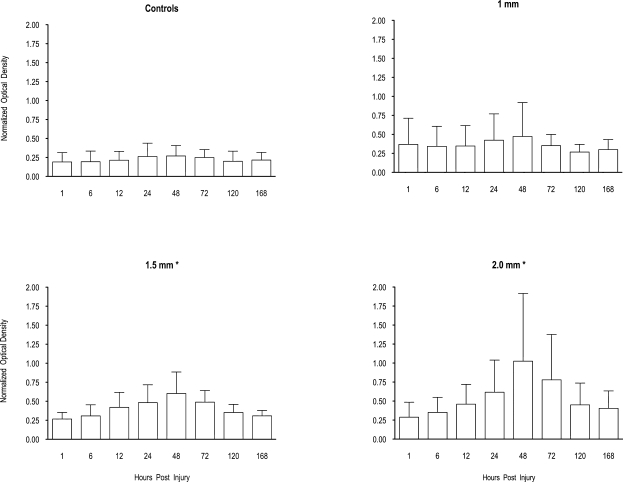
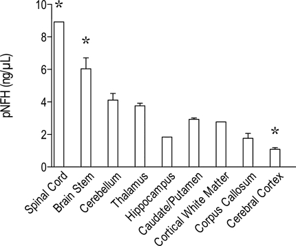
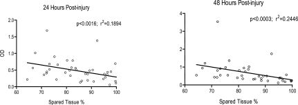
The detection of neuron-specific proteins in blood might allow quantification of the degree of neuropathology in experimental and clinical contexts. We have been studying a novel blood biomarker of axonal injury, the heavily phosphorylated axonal form of the high molecular weight neurofilament subunit NF-H (pNF-H). We hypothesized that this protein would be released from damaged and degenerating neurons following experimental traumatic brain injury (TBI) in amounts large enough to allow its detection in blood and that the levels detected would reflect the degree of injury severity. An enzyme-linked immunosorbent assay (ELISA) capture assay capable of detecting nanogram amounts of pNF-H was used to test blood of rats subjected to experimental TBI using a controlled cortical impact (CCI) device. Animals were subjected to a mild (1.0 mm), moderate (1.5 mm), or severe (2.0 mm) cortical contusion, and blood samples were taken at defined times post-injury. The assay detected the presence of pNF-H as early as 6 h post-injury; levels peaked at 24-48 h, and then slowly decreased to baseline over several days post-injury. No signal above baseline was detectable in control animals. Analysis of variance (ANOVA) showed a significant effect of lesion severity, and post hoc analysis revealed that animals given a moderate and severe contusion showed higher levels of blood pNF-H than controls. In addition, the peak levels of pNF-H detected at both 24 and 48 h post-injury correlated with the degree of injury as determined by volumetric analysis of spared cortical tissue. Relative amounts of pNF-H were also determined in different areas of the central nervous system (CNS) and were found to be highest in regions containing large-diameter axons, including spinal cord and brainstem, and lowest in the cerebral cortex and hippocampus. These findings suggest that the measurement of blood levels of pNF-H is a convenient method for assessing neuropathology following TBI.
Figures
References
-
- Anderson R.E. Hansson L.O. Nilsson O. Dijlai-Merzoug R. Settergren G. High serum S100B levels for trauma patients without head injuries. Neurosurgery. 2001;48:1255–1260. - PubMed
-
- Baldwin S.A. Gibson T. Callihan C.T. Sullivan P.G. Palmer E. Scheff S.W. Neuronal cell loss in the CA3 subfield of the hippocampus following cortical contusion utilizing the optical disector method for cell counting. J. Neurotrauma. 1997;14:385–398. - PubMed
-
- Baldwin S.A. Scheff S.W. Intermediate filament change in astrocytes following mild cortical contusion. Glia. 1996;16:266–275. - PubMed
-
- Baskaya M.K. Rao A.M. Dogan A. Donaldson D. Dempsey R.J. The biphasic opening of the blood-brain barrier in the cortex and hippocampus after traumatic brain injury in rats. Neurosci. Lett. 1997;226:33–36. - PubMed
-
- Beer R. Franz G. Srinivasan A. Hayes R.L. Pike B.R. Newcomb J.K. Zhao X. Schmutzhard E. Poewe W. Kampfl Et A. Temporal profile and cell subtype distribution of activated caspase-3 following experimental traumatic brain injury. J. Neurochem. 2000;75:1264–1273. - PubMed
Publication types
MeSH terms
Substances
Grants and funding
LinkOut - more resources
Full Text Sources
Other Literature Sources
