

Effects of host temperature and gastric and duodenal environments on microsporidia spore germination and infectivity of intestinal epithelial cells
- PMID: 18751726
- PMCID: PMC2737319
- DOI: 10.1007/s00436-008-1156-4
Effects of host temperature and gastric and duodenal environments on microsporidia spore germination and infectivity of intestinal epithelial cells
Abstract
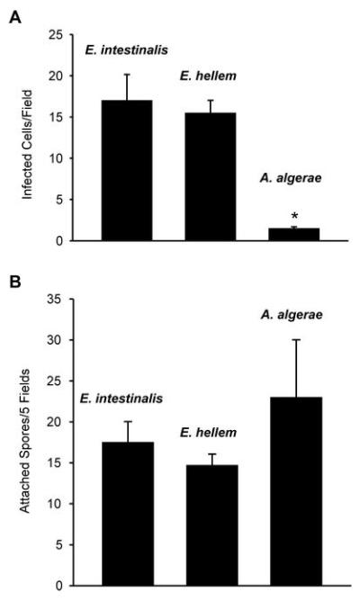
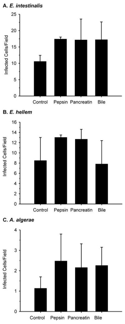
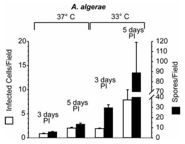
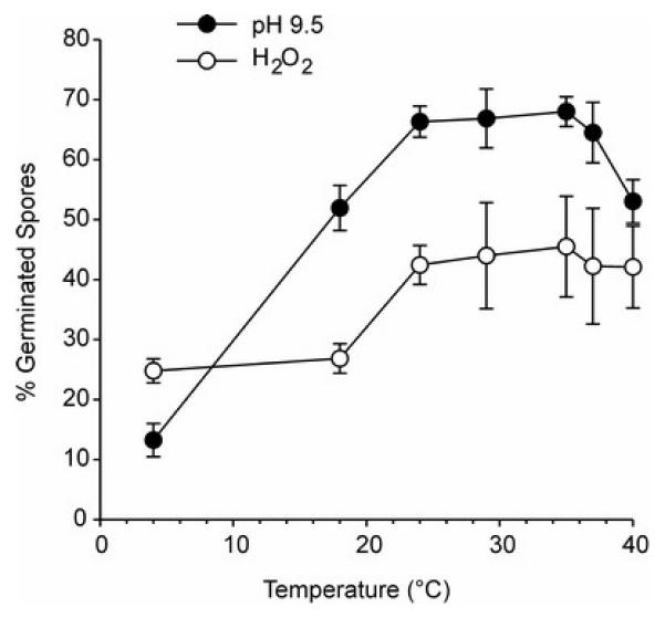
Approximately 14 of the more than 1,000 species of microsporidia infect humans, only two of which, Enterocytozoon bieneusi and Encephalitozoon intestinalis, cause intestinal microsporidiosis. Clinical isolates of three microsporidia species, E. intestinalis, Encephalitozoon hellem, and the insect parasite, Anncaliia (Brachiola, Nosema) algerae were used in a spore germination assay, and enterocyte attachment and infection assays were performed to model the potential roles of gastric and duodenal environments and host temperature in determining why only one of these microsporidia species causes intestinal microsporidiosis. Enterocyte infection with A. algerae spores was 10% that of the Encephalitozoon species, a difference not attributable to differences in spore attachment to host cells. Prior spore treatment with pepsin in HCl, pancreatic enzymes, or ox bile did not inhibit germination or enterocyte infection by the three microsporidia species. While the Encephalitozoon species differentiated to mature spores within 3 days, the time taken for many enterocytes to turn over, A. algerae took 3-5 days to produce mature spores, near the upper limit for enterocyte turnover in vivo. Thus, host temperature may contribute to A. algerae not causing human intestinal microsporidiosis, but none of the factors tested account for the inability of E. hellem to cause such an infection.
Figures
Similar articles
-
A role for antimicrobial peptides in intestinal microsporidiosis.Parasitology. 2009 Feb;136(2):175-81. doi: 10.1017/S0031182008005313. Epub 2008 Dec 12. Parasitology. 2009. PMID: 19079820 Free PMC article.
-
[Animal reservoirs of human virulent microsporidian species].Wiad Parazytol. 2009;55(1):63-5. Wiad Parazytol. 2009. PMID: 19579789 Polish.
-
[Investigation of Microsporidia prevalence with calcofluor white and uvitex 2B chemiluminescence staining methods and molecular analysis of species in diarrheal patients].Mikrobiyol Bul. 2018 Oct;52(4):401-412. doi: 10.5578/mb.67363. Mikrobiyol Bul. 2018. PMID: 30522425 Turkish.
-
In vitro cultivation of microsporidia of clinical importance.Clin Microbiol Rev. 2002 Jul;15(3):401-13. doi: 10.1128/CMR.15.3.401-413.2002. Clin Microbiol Rev. 2002. PMID: 12097248 Free PMC article. Review.
-
[Microsporidia: general characteristics, infections and laboratory diagnosis].Mikrobiyol Bul. 2005 Oct;39(4):513-22. Mikrobiyol Bul. 2005. PMID: 16544554 Review. Turkish.
Cited by
-
Factors That Determine Microsporidia Infection and Host Specificity.Exp Suppl. 2022;114:91-114. doi: 10.1007/978-3-030-93306-7_4. Exp Suppl. 2022. PMID: 35544000 Review.
-
In vitro growth of microsporidia Anncaliia algerae in cell lines from warm water fish.In Vitro Cell Dev Biol Anim. 2011 Feb;47(2):104-13. doi: 10.1007/s11626-010-9366-3. Epub 2010 Nov 18. In Vitro Cell Dev Biol Anim. 2011. PMID: 21086187
-
High temperatures and low humidity promote the occurrence of microsporidians (Microsporidia) in mosquitoes (Culicidae).Parasit Vectors. 2024 Apr 11;17(1):187. doi: 10.1186/s13071-024-06254-0. Parasit Vectors. 2024. PMID: 38605410 Free PMC article.
-
Invasion of Host Cells by Microsporidia.Front Microbiol. 2020 Feb 18;11:172. doi: 10.3389/fmicb.2020.00172. eCollection 2020. Front Microbiol. 2020. PMID: 32132983 Free PMC article. Review.
-
A role for antimicrobial peptides in intestinal microsporidiosis.Parasitology. 2009 Feb;136(2):175-81. doi: 10.1017/S0031182008005313. Epub 2008 Dec 12. Parasitology. 2009. PMID: 19079820 Free PMC article.
References
-
- Avery SW, Undeen AH. The isolation of microsporidia and other pathogens from concentrated ditch water. J Am Mosq Control Assoc. 1987;3:54–58. - PubMed
-
- Cotte L, Rabodonirina M, Chapuis F, Bailly F, Bissuel F, Raynal C, Gelas P, Persat F, Piens M-A, Trepo C. Waterborne outbreak of intestinal microsporidiosis in persons with and without human immunodeficiency virus infection. J Infect Dis. 1999;180:2003–2008. - PubMed
-
- Didier ES. Microsporidiosis: and emerging and opportunistic infection in humans and animals. Acta Trop. 2005;94:61–76. - PubMed
Publication types
MeSH terms
Grants and funding
LinkOut - more resources
Full Text Sources
