. 2007 Feb;3(1):13-29.
doi: 10.1007/s11420-006-9025-0.

The mechanics of external fixation

Affiliations

The mechanics of external fixation

Austin T Fragomen et al. HSS J. 2007 Feb.

Abstract

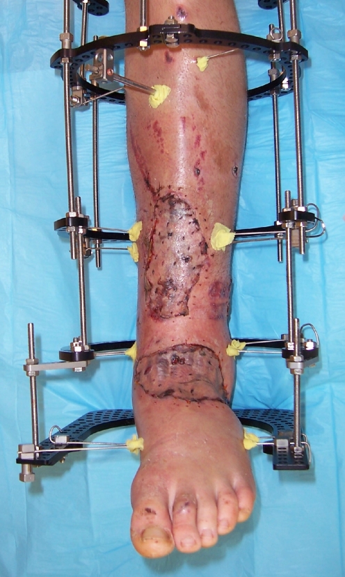
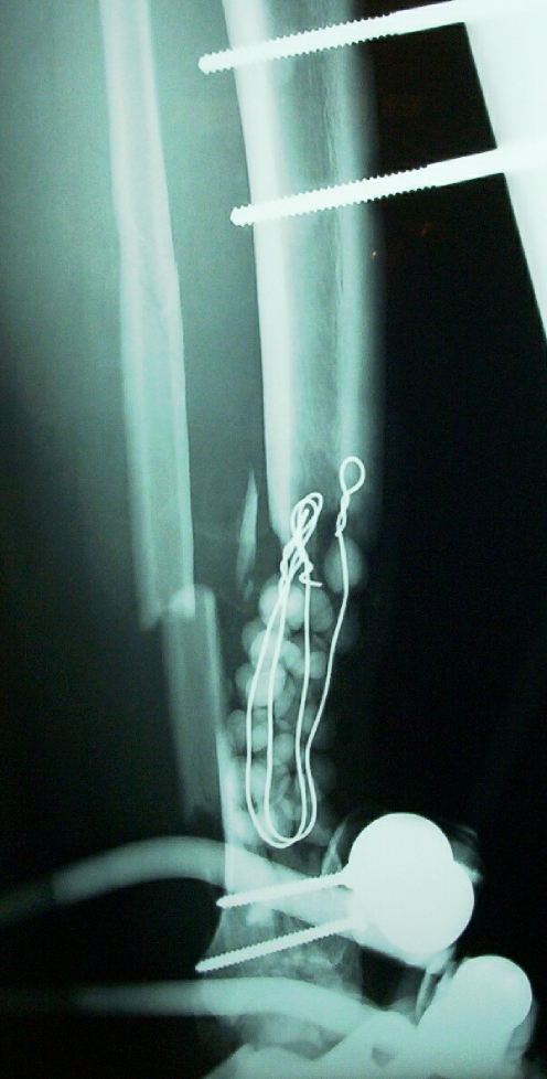
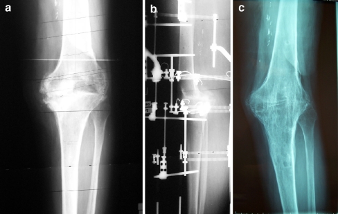
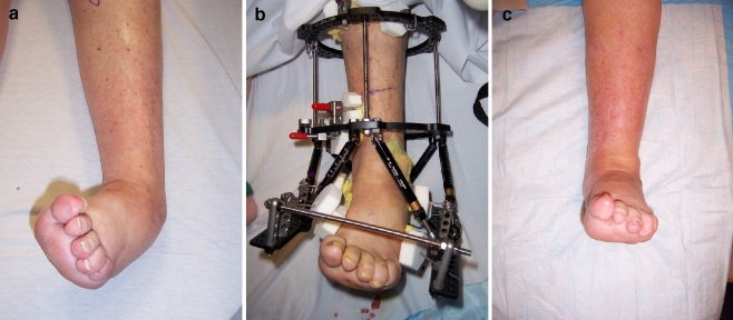
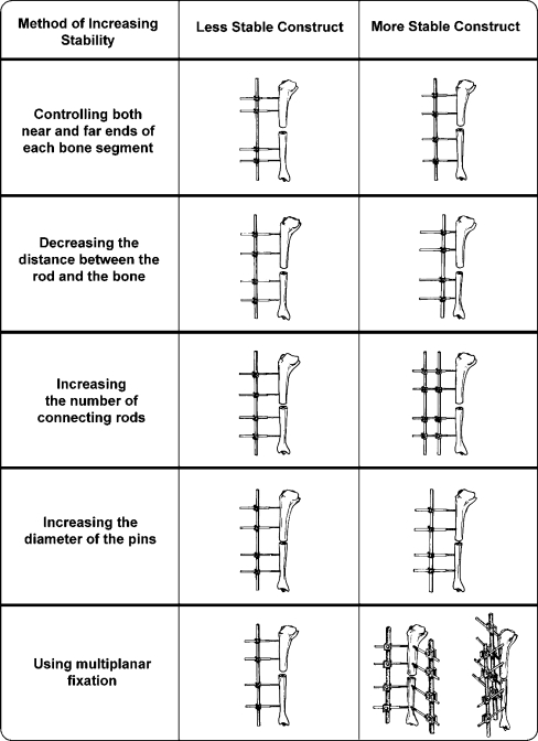
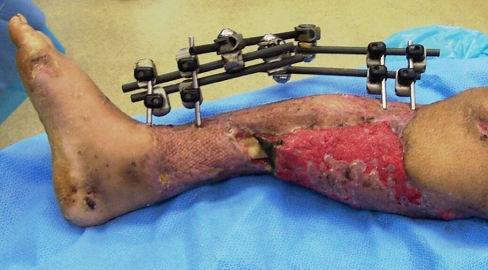
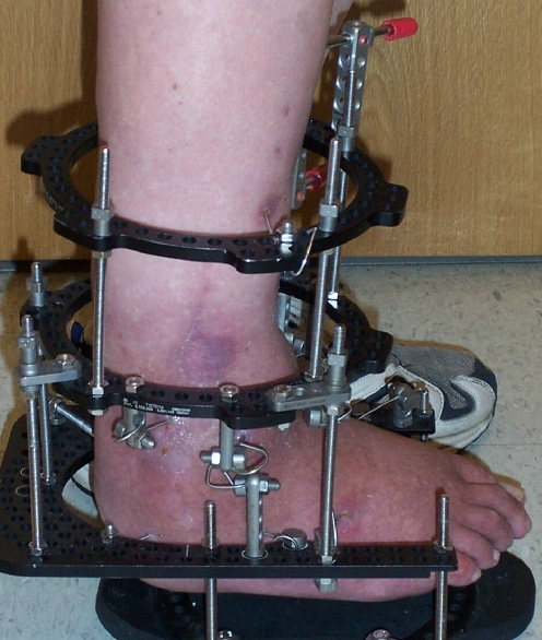
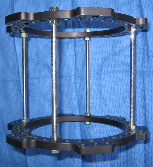
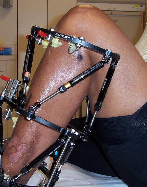
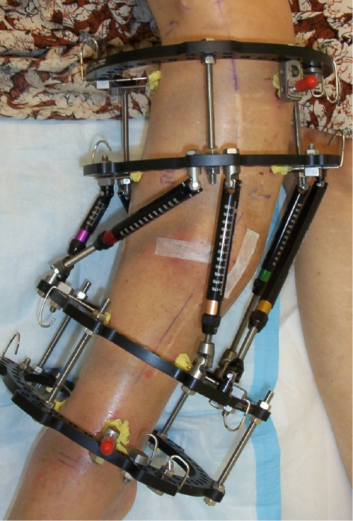
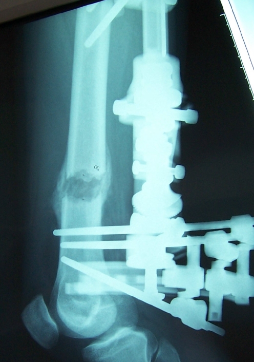
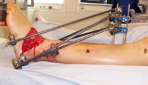
External fixation has evolved from being used primarily as a last resort fixation method to becoming a main stream technique used to treat a myriad of bone and soft tissue pathologies. Techniques in limb reconstruction continue to advance largely as a result of the use of these external devices. A thorough understanding of the biomechanical principles of external fixation is useful for all orthopedic surgeons as most will have to occasionally mount a fixator throughout their career. In this review, various types of external fixators and their common clinical applications are described with a focus on unilateral and circular frames. The biomechanical principles that govern bony and fixator stability are reviewed as well as the recommended techniques for applying external fixators to maximize stability. Additionally, we have illustrated methods for managing patients while they are in the external frames to facilitate function and shorten treatment duration.


Figures

Fig. 1
Fig. 1
In this case of significant lower extremity trauma, the Ilizarov frame provided stability to the fracture, protected the skin grafts, and allowed access to the involved soft tissues. These percutaneous K-wires cause minimal disruption of the blood supply and soft tissues through the zone of injury
Fig. 2
Fig. 2
Trauma type pin-to-bar fixator used to help eradicate infection in a septic tibial nonunion
Fig. 3
Fig. 3
(a) This patient failed revision knee replacement surgery for recurrent infection. (b) The Ilizarov fixator was applied for knee fusion.(c) Healed knee fusion
Fig. 4
Fig. 4
(a) This 82 year old woman with diabetes mellitus and advanced peripheral vascular disease had an aseptic tibial nonunion. (b) She was treated with circular fixation and gradual correction of the deformity without open repair of the nonunion. (c) Healed nonunion. The patient was in the frame for 4 months
Fig. 5
Fig. 5
(a) This severe varus deformity was corrected through an ankle fusion. (b) A gradual correction approach was taken. (c) Final result after frame removal
Fig. 6
Fig. 6
Unilateral external fixator stability diagram
Fig. 7
Fig. 7
The EBI MAC frame is quite rigid and allows for the correction of multiplanar deformities
Fig. 8
Fig. 8
This unilateral fixator has gained stability by connecting a second bar to each pair of half pins
Fig. 9
Fig. 9
This ankle fusion frame combines the classic Ilizarov assembly with the modern aluminum Taylor Spatial rings. This makes the frame more modular as at any time the connecting rods could be exchanged for spatial struts. If a proximal lengthening is planned then existing rings are ready to accept struts as well
Fig. 10
Fig. 10
Taylor spatial rings connected with 10 mm threaded connecting rods
Fig. 11
Fig. 11
The 2/3 ring is used around the knee joint to allow improved joint flexion. Rehabilitation begins during treatment
Fig. 12
Fig. 12
This proximal femoral osteotomy was stabilized with a two Ilizarov arches
Fig. 13
Fig. 13
Despite the use of large rings this patient’s leg swelled to the point of impinging on the middle ring. The skin was observed closely but no intervention was necessary
Fig. 14
Fig. 14
This lengthening frame incorporates a middle ring that has no bony attachment. This “dummy ring” serves to improve fixator stability over long distances
Fig. 15
Fig. 15
This severely deformed leg requires the use of two stable ring blocks for correction. In this example each ring block consists of 2 rings with 4 points of fixation. Each ring block is aligned with the axis of its respective bone segment. This crooked frame will be straight when the correction is completed
Fig. 16
Fig. 16
Femoral lengthening performed with a unilateral fixator
Fig. 17
Fig. 17
This open pilon fracture was stabilized with an ankle spanning simple construct
Fig. 18
Fig. 18
This construct emphasizes the freedom that these frames allow for pin placement and incorporation into the frame
Fig. 19
Fig. 19
(a) This temporary two ring frame provides provisional stability and allows access for soft tissue management. (b) Definitive bony treatment follows, in this instance with a more stable bone transport set up
Fig. 20
Fig. 20
A typical proximal tibial ring block consists of one ring with 4 points of fixation. Although the two wires have a narrow crossing angle (25 degrees), the 2 half pins prevent any sliding of the ring in the coronal plane. These two wires provide excellent resistance to medial–lateral bending forces
Fig. 21
Fig. 21
Typical hydroxyapatite (HA) coated, 5–to–6 mm tapered half pin. This would be used in the adult tibia, femur and humerus
Fig. 22
Fig. 22
This knee spanning design provides suboptimal stability for ambulation as a minimal number of pins were used, the pins are clustered close together, and there is poor control over sagittal motion
Fig. 23
Fig. 23
(a) Persistent knee flexion contracture after total knee replacement surgery was treated with Ilizarov method using hinges. (b) Full knee extension was accomplished safely using this method
Fig. 24
Fig. 24
The Taylor Spatial Frame has the unique ability to apply compression to a fracture in an oblique plane. By telling the computer to “shorten” (axially compress) and “posteriorly translate” the distal fragment, a net vector of compression is created in an oblique plane perpendicular to the fracture plane

References

    1. {'text': '', 'ref_index': 1, 'ids': [{'type': 'PubMed', 'value': '2647333', 'is_inner': True, 'url': 'https://pubmed.ncbi.nlm.nih.gov/2647333/'}]}
    2. Behrens F (1989) General theories and principles of external fixation. Clin Orthop 241:15–23 - PubMed
    1. {'text': '', 'ref_index': 1, 'ids': [{'type': 'DOI', 'value': '10.1097/00003086-199908000-00027', 'is_inner': False, 'url': 'https://doi.org/10.1097/00003086-199908000-00027'}, {'type': 'PubMed', 'value': '10627706', 'is_inner': True, 'url': 'https://pubmed.ncbi.nlm.nih.gov/10627706/'}]}
    2. Claes L, Heitemeyer U, Krischak G, et al. (1999) Fixation technique influences osteogenesis of comminuted fractures. Clin Orthop 365:221–229 - PubMed
    1. {'text': '', 'ref_index': 1, 'ids': [{'type': 'DOI', 'value': '10.1097/00005131-200503000-00004', 'is_inner': False, 'url': 'https://doi.org/10.1097/00005131-200503000-00004'}, {'type': 'PubMed', 'value': '15758670', 'is_inner': True, 'url': 'https://pubmed.ncbi.nlm.nih.gov/15758670/'}]}
    2. Rozbruch SR, Ilizarov S, Blyakher A (2005) Knee arthrodesis with simultaneous lengthening using the Ilizarov method. J Orthop Trauma 19:171–179 - PubMed
    1. {'text': '', 'ref_index': 1, 'ids': [{'type': 'PubMed', 'value': '3514629', 'is_inner': True, 'url': 'https://pubmed.ncbi.nlm.nih.gov/3514629/'}]}
    2. Behrens F, Searls K (1986) External fixation of the tibia. Basic concepts and prospective evaluation. J Bone Joint Surg Br 62:246–254 - PubMed
    1. {'text': '', 'ref_index': 1, 'ids': [{'type': 'PubMed', 'value': '6883844', 'is_inner': True, 'url': 'https://pubmed.ncbi.nlm.nih.gov/6883844/'}]}
    2. Behrens F, Johnson WD, Koch TW, Kovacevic N (1983) Bending stiffness of unilateral and bilateral fixation frames. Clin Orthop 178:103–110 - PubMed