

doi: 10.1007/s11420-005-0129-8.
Haglund's syndrome: diagnosis and treatment using sonography
Affiliations
- PMID: 18751843
- PMCID: PMC2504114
- DOI: 10.1007/s11420-005-0129-8
Item in Clipboard
Haglund's syndrome: diagnosis and treatment using sonography
HSS J.
2006 Feb.
Abstract
Haglund's syndrome is a cause of retrocalcaneal pain. The clinical diagnosis of Haglund's syndrome is often confusing as the clinical picture may mimic other causes of hindfoot pain such as isolated retrocalcaneal bursitis or hindfoot involvement from more systemic arthropathies such as Reiter's syndrome or rheumatoid arthritis. With the increasing frequency of employing sonography as a diagnostic tool in the evaluation of foot and ankle pathology, recognition of the sonographic appearance of Haglund's complex is important. We report a case of Haglund's syndrome diagnosed and treated with sonography.
Figures
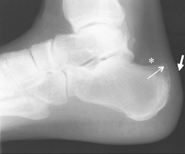
Lateral standing radiograph of the hindfoot demonstrates a prominent posterosuperior osseous calcaneal protuberance (arrow) with a vague, cloudy density in the deep retrocalcaneal bursa (*) consistent with retrocalcaneal bursitis. Superficial tendoAchilles bursitis is demonstrated by convexity of the soft tissues (short thick arrow) and an ill-defined Achilles tendon at its insertion consistent with inflammation
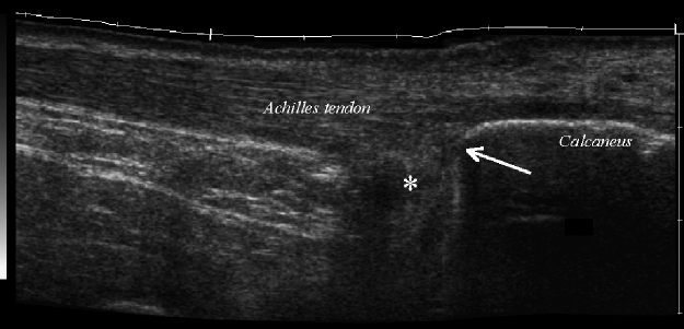
Longitudinal extended field of view sonographic image of the hindfoot demonstrates the prominent osseous protuberance at the posterosuperior margin of the calcaneus (arrow) and hypoechoic distention of the retrocalcaneal bursa (*). Thickening of the superficial tendon Achilles bursa is also present
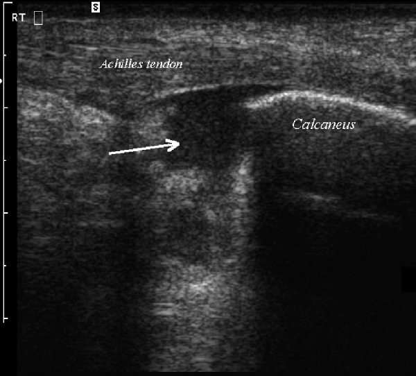
Longitudinal sonogram of the hindfoot after ultrasound guided retrocalcaneal bursal injection demonstrates hypoechoic material with internal low levels echoes distending the bursa, representing the injected steroid/anesthetic material (arrow)
References
-
- Haglund P. Beitrag zur Klinik der Achillessehne. Z Orthop Chir. 1927;49:49–58.
-
- Pavlov H, Heneghan MA, Hersh A, Goldman AB, Vigorita V. The Haglund syndrome: initial and differential diagnosis. Radiology. 1982;144(1):83–88. - PubMed
-
- Olivieri I, Barozzi L, Padula A, DeMatteis M, Pierro A, Cantini F, Salvarani C, Pavlica P. Retrocalcaneal bursitis in spondyloarthropathy: assessment by ultrasonography and magnetic resonance imaging. J Rheumatol. 1998;25(7):1352–1357. - PubMed
-
- Stephens MM. Haglund's deformity and retrocalcaneal bursitis. Orthop Clin North Am. 1994;25(1):41–46. - PubMed
-
- Heneghan MA, Pavlov H. The Haglund painful heel syndrome: experimental investigation of cause and therapeutic implications. Clin Orthop. 1984;187:228–234. - PubMed
LinkOut - more resources
Full Text Sources
