. 2008 Sep;48(3):841-52.
doi: 10.1002/hep.22401.

Natural killer T cell dysfunction in CD39-null mice protects against concanavalin A-induced hepatitis

Affiliations

Natural killer T cell dysfunction in CD39-null mice protects against concanavalin A-induced hepatitis

Guido Beldi et al. Hepatology. 2008 Sep.

Erratum in

  • Hepatology. 2009 Jul;50(1):331

Abstract

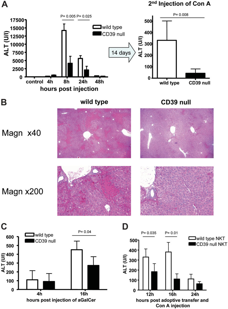
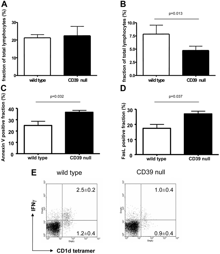
Concanavalin A (Con A)-induced injury is an established natural killer T (NKT) cell-mediated model of inflammation that has been used in studies of immune liver disease. Extracellular nucleotides, such as adenosine triphosphate, are released by Con A-stimulated cells and bind to specific purinergic type 2 receptors to modulate immune activation responses. Levels of extracellular nucleotides are in turn closely regulated by ectonucleotidases, such as CD39/NTPDase1. Effects of extracellular nucleotides and CD39 on NKT cell activation and upon hepatic inflammation have been largely unexplored to date. Here, we show that NKT cells express both CD39 and CD73/ecto-5'-nucleotidase and can therefore generate adenosine from extracellular nucleotides, whereas natural killer cells do not express CD73. In vivo, mice null for CD39 are protected from Con A-induced liver injury and show substantively lower serum levels of interleukin-4 and interferon-gamma when compared with matched wild-type mice. Numbers of hepatic NKT cells are significantly decreased in CD39 null mice after Con A administration. Hepatic NKT cells express most P2X and P2Y receptors; exceptions include P2X3 and P2Y11. Heightened levels of apoptosis of CD39 null NKT cells in vivo and in vitro appear to be driven by unimpeded activation of the P2X7 receptor.

Conclusion: CD39 and CD73 are novel phenotypic markers of NKT cells. In turn, CD39 expression [corrected] modulates nucleotide-mediated cytokine production by, and limits apoptosis of, hepatic NKT cells. Deletion of CD39 is protective in [corrected] Con A-induced hepatitis. This study illustrates a [corrected] role for purinergic signaling in NKT-mediated mechanisms that result in liver immune injury.


Conflict of interest statement

Potential conflict of interest: Nothing to report.

Figures

Fig. 1
Fig. 1
NKT cell populations in wild-type and CD39-null mice. (A) Characterization of isolated liver MNCs from wild-type mice and CD39-null mice. Basal levels of hepatic NK1.1+CD3+, NK1.1+CD3−, and αGalCer-loaded CD1d tetramer+ cells are shown. (B) All hepatic NK1.1+ and αGalCer-loaded CD1d tetramer+ cells expressed CD39. Data are representative of four independent experiments with similar results.
Fig. 2
Fig. 2
CD73 expression is confined to NKT and T cells. Expression patterns of CD73/ecto-5′-ectonucleotidase on NK1.1+CD3− (NK), NK1.1+CD3+ (NKT), and NK1.1−CD3+ (T cell) subsets of liver MNCs and splenocytes are shown in two sections. (A) NKT and T cells in liver MNCs express CD73; NK cells do not. (B) Splenocyte populations were examined and, as in the liver, show that the majority of NK cells do not express CD73. Data are representative of three independent experiments with similar results.
Fig. 3
Fig. 3
Biochemical analysis of NTPDase activity of hepatic MNCs and sorted NKT cells from wild-type and CD39-null mice. (A) Time course of [3H]ATP hydrolysis and formation of its dephosphorylated 3H-metabolites by liver MNCs from wild-type (left panel) and CD39-null (right panel) mice (n = 4 each). The ordinate shows relative contents of [3H]-nucleotides/adenosine expressed as percentage of initial concentrations. (B) Analysis of the products of [14C]ADP hydrolysis by purified hepatic NKT cells from wild-type and CD39-null mice (n = 6). Extracellular nucleotides are efficiently hydrolyzed to AMP and adenosine by wild-type cells, whereas CD39-null cells showed delayed hydrolysis of nucleotides and limited production of adenosine.
Fig. 4
Fig. 4
Immune hepatitis model and CD39. (A). CD39-null mice were significantly protected from Con A–induced hepatitis (15 mg/kg) at various time points (n = 5 per time point) after injection. Minimal injury was observed in CD39-null mice after a second injection of 20 mg/kg Con A (n = 5). (B) Representative histological sections 16 hours after initial injection of Con A are shown. Magnifications are ×40 and ×200. (C) Liver injury was assessed after injection of 100 ng αGalCer per mouse at various time points (n = 4 per time point). CD39-null mice showed significantly reduced injury as assessed by ALT levels. (D) Adoptive transfer of wild-type and CD39-null NKT cells was performed in CD1d-null mice with concurrent injection of 15 mg/kg Con A (n = 4). NKT null for CD39 failed to induce significant liver injury 12 and 16 hours after injection. Data are presented as the mean ± standard deviation.
Fig. 5
Fig. 5
NKT cell–associated cytokine levels in vivo and in vitro. (A) Serum levels of IL-4 and IFN-γ determined after Con A injection were significantly decreased in CD39-null mice. (B) Secretion of IL-4 and IFN-γ by NKT cells in response to αGalCer. Liver MNCs were stimulated with αGalCer or Con A for 24 hours in vitro. The secretion of IL-4 and IFN-γ was significantly less from CD39-null cells. (C) Secretion of IL-4 and IFN-γ by NKT cells in response to αGalCer-primed dendritic cells. Sorted splenic dendritic cells were loaded with αGalCer for 2 hours and subsequently incubated with liver MNCs (as designated on horizontal axis) in vitro for 24 hours. The secretion of IL-4 was significantly less in MNCs null for CD39, whereas in the presence of dendritic cells the secretion of IFN-γ was increased over wild-type controls (*P < 0.05). The differential expression of CD39 on splenic dendritic cells in coculture experiments did not alter the cytokine secretion. Data are given as the mean ± standard deviation of at least four animals per group (A) and/or representative of three independent experiments (B,C).
Fig. 6
Fig. 6
Apoptosis of NKT cells in Con A–induced liver injury. (A) Under basal conditions the fraction of hepatic invariant NKT cells is not different between wild-type mice and CD39-null mice. (B) Sixty minutes after injection of Con A, the NKT cell count was significantly decreased in CD39-null livers (n = 4). (C,D) Fractions of (C) Annexin V–positive and (D) FasL-positive NKT cells were increased in mice null for CD39 compared with wild-type mice. Data are representative of at least three experiments with similar results. (E) Expression of fractions of intracellular IFN-γ–positive and IFN-γ–negative NKT cells (αGalCer-loaded CD1d tetramer+ cells) of liver MNCs after Con A–induced hepatitis were assessed via fluorescence-activated cell sorting. The number of NKT cells and the fraction of IFN-γ–positive NKT cells was decreased in CD39-null mice compared with wild-type cells (P = 0.005). Data are given as the mean ± standard deviation of four independent experiments.
Fig. 7
Fig. 7
NKT cells express multiple P2 receptors. (A) Analysis of the messenger RNA expression of P2Y and P2X receptors on hepatic NKT cells (NK1.1+, CD3+). RT-PCR revealed that all of the known P2 receptors, with the exception of P2Y11 and P2X3, were found on quiescent NKT cells. (B) The expression of P2 receptors was assessed using real-time RT-PCR. The CD4+ and CD4− subsets of isolated hepatic, splenic, and thymic NKT (CD3+, NK1.1+) cells were analyzed separately.
Fig. 8
Fig. 8
Induction of NKT apoptosis by extracellular ATP in vitro. Isolated liver MNCs from wild-type and CD39-null mice were incubated with various levels of ATP, and the extent of apoptosis was then determined. (A) The fraction of Annexin V–positive NKT cells is given as green histograms for wild-type cells and CD39-null cells. Extracellular ATP resulted in significantly increased expression of Annexin V on hepatic NKT cells null for CD39 compared with wild-type NKT cells (analysis of variance, P = 0.02). (B) The fraction of NKT versus the total number of MNCs reveals a dose-dependent decrease of NKT cells with increasing doses of ATP (analysis of variance, P = 0.015). (C) Administration of oxidized ATP (a P2X7 antagonist) was associated with a relative decrease of Annexin V–positive NKT cells (*P < 0.05 wild-type versus CD39-null). (A,C) Representative results of three experiments. (B) Mean and standard error of the mean from five experiments.

References

    1. Tiegs G, Hentschel J, Wendel A. A T cell-dependent experimental liver injury in mice inducible by concanavalin A. J Clin Invest. 1992;90:196–203. - PMC - PubMed
    1. Schumann J, Wolf D, Pahl A, Brune K, Papadopoulos T, van Rooijen N, et al. Importance of Kupffer cells for T-cell-dependent liver injury in mice. Am J Pathol. 2000;157:1671–1683. - PMC - PubMed
    1. Exley MA, Koziel MJ. To be or not to be NKT: natural killer T cells in the liver. HEPATOLOGY. 2004;40:1033–1040. - PubMed
    1. Takeda K, Hayakawa Y, Van Kaer L, Matsuda H, Yagita H, Okumura K. Critical contribution of liver natural killer T cells to a murine model of hepatitis. Proc Natl Acad Sci U S A. 2000;97:5498–5503. - PMC - PubMed
    1. Toyabe S, Seki S, Iiai T, Takeda K, Shirai K, Watanabe H, et al. Requirement of IL-4 and liver NK1+ T cells for concanavalin A-induced hepatic injury in mice. J Immunol. 1997;159:1537–1542. - PubMed

Publication types

MeSH terms