. 2008 Nov 7;283(45):31246-55.
doi: 10.1074/jbc.M806085200. Epub 2008 Aug 27.

DUSP6 (MKP3) null mice show enhanced ERK1/2 phosphorylation at baseline and increased myocyte proliferation in the heart affecting disease susceptibility

Affiliations

DUSP6 (MKP3) null mice show enhanced ERK1/2 phosphorylation at baseline and increased myocyte proliferation in the heart affecting disease susceptibility

Marjorie Maillet et al. J Biol Chem. .

Abstract

The strength and duration of mitogen-activated protein kinase signaling is regulated through phosphorylation and dephosphorylation by dedicated dual-specificity kinases and phosphatases, respectively. Here we investigated the physiological role that extracellular signal-regulated kinases 1/2 (ERK1/2) dephosphorylation plays in vivo through targeted disruption of the gene encoding dual-specificity phosphatase 6 (Dusp6) in the mouse. Dusp6(-/-) mice, which were viable, fertile, and otherwise overtly normal, showed an increase in basal ERK1/2 phosphorylation in the heart, spleen, kidney, brain, and fibroblasts, but no change in ERK5, p38, or c-Jun N-terminal kinases activation. However, loss of Dusp6 did not increase or prolong ERK1/2 activation after stimulation, suggesting that its function is more dedicated to basal ERK1/2 signaling tone. In-depth analysis of the physiological effect associated with increased baseline ERK1/2 signaling was performed in cultured mouse embryonic fibroblasts (MEFs) and the heart. Interestingly, mice lacking Dusp6 had larger hearts at every age examined, which was associated with greater rates of myocyte proliferation during embryonic development and in the early postnatal period, resulting in cardiac hypercellularity. This increase in myocyte content in the heart was protective against decompensation and hypertrophic cardiomyopathy following long term pressure overload and myocardial infarction injury in adult mice. Dusp6(-/-) MEFs also showed reduced apoptosis rates compared with wild-type MEFs. These results demonstrate that ERK1/2 signaling is physiologically restrained by DUSP6 in coordinating cellular development and survival characteristics, directly impacting disease-responsiveness in adulthood.


Figures
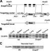
FIGURE 1.
FIGURE 1.
Gene targeting of Dusp6. A, schematic of the Dusp6 genetic locus in the mouse and the targeting vector that was used to replace exons 1-3 in embryonic stem cells. B, Western blot analysis for DUSP6 protein generated from the indicated tissues from wild-type (Wt) or Dusp6-/- neonates. C, Western blot analysis for DUSP6 protein generated from hearts of Wt or Dusp6-/- neonates (3 days old).
FIGURE 2.
FIGURE 2.
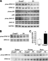
Deletion of Dusp6 augments baseline ERK1/2 phosphorylation. A, Western blots for MAPK and phospho-MAPK proteins from cultured MEFs derived from Wt or Dusp6-/- embryos. Glyceraldehyde-3-phosphate dehydrogenase (GAPDH) was run as a loading control. B, Western blotting of baseline phospho-ERK1/2 and total ERK1/2 from 1-month-old hearts of Wt and Dusp6-/- mice (C) and associated quantitation. D, Western blotting for baseline phospho-ERK1/2 and total ERK1/2 from the spleen, kidney, and brain of 1-month-old Wt and Dusp6-/- mice. Tissue from at least four separate animals was examined in each group. Data are representative of three independent experiments.
FIGURE 3.
FIGURE 3.
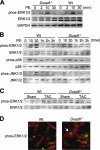
ERK1/2 phosphorylation kinetics are unaffected in the absence of Dusp6. A, Western blot for phospho-ERK1/2 and total ERK1/2 from cultured Wt or Dusp6-/- MEFs stimulated with PE for the indicated time points in minutes under serum-starved conditions. B, Western blotting for phospho-MAPKs and total MAPKs from the hearts of 2-month-old Wt and Dusp6-/- mice injected with PE systemically (10 mg/kg) for the indicated period of time in minutes or hours (h). C, Western blotting for phospho-ERK1/2 or total ERK1/2 from the hearts of 2-month-old Wt or Dusp6-/- mice subjected to TAC-induced pressure overload or sham surgical procedure and harvested 2 weeks later. D, Dusp6-/- and Wt MEFs in serum-free media after PE stimulation were stained for phospho-ERK1/2 (green) and actin (red). The arrow shows perinuclear phospho-ERK1/2. Data shown throughout this figure are representative of at least three independent experiments.
FIGURE 4.
FIGURE 4.
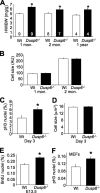
Greater proliferation of myocytes in the hearts of Dusp6-/- mice. A, quantitation of heart weight normalized to body weight (HW/BW) at 1, 2 and 12 months of age in Wt and Dusp6-/- mice (n = 4 or more each). *, p < 0.05 versus Wt control at the same age. B, quantitation of myocyte surface area from histological heart sections at 1 month of age or total surface area of individual myocytes isolated from hearts of 2-month-old Wt or Dusp6-/- mice. Wt values were normalized to 100 arbitrary units (AU). C, immunohistochemical quantitation of phospho-histone H3 levels in myocytes from 3 day-old Wt and Dusp6-/- mice (n = 2000 nuclei counted each);D, with corresponding cell cross-sectionalareas. *, p <0.05 versus Wt. E, immunohistochemical quantitation of BrdUrd incorporation in the hearts of day 13.5 embryos from Wt and Dusp6-/- mice (n = 4000-6000 nuclei counted from two different embryos). *, p < 0.05 versus Wt. F, phospho-histone H3 levels measured by immunocytochemistry in cultures of Wt and Dusp6-/- MEFs expressed as a percentage of total nuclei (n = 500 nuclei counted for each). *, p < 0.05 versus Wt.
FIGURE 5.
FIGURE 5.
Examination of the cardiac hypertrophic response in Dusp6-/- mice following stimulation. A, quantitation of HW/BW in 2-month-old Wt or Dusp6-/- mice implanted with control (saline) or AngII/PE containing Alzet mini-pumps (432 μg/kg/day, 100 mg/kg/day, respectively) for 2 weeks (n = 6 or more mice in each group). *, p < 0.05 versus Wt saline; NS, not significant. B, quantitation of HW/BW in 2-month-old Wt or Dusp6-/- mice implanted with control (saline) or isoprenaline (Iso) containing Alzet mini-pumps (60 mg/kg/day) for 2 weeks (n = 6 or more mice in each group). *, p < 0.05 versus Wt saline; #, p < 0.05 versus Wt isoprenaline; NS, not significant. C, quantitation of HW/BW in 2-month-old Wt or Dusp6-/- mice subjected to TAC for 8 weeks (n = 8 Wt saline, 6 Dusp6-/- saline, 13 Wt TAC, and 11 Dusp6-/- TAC). *, p < 0.05 versus Wt sham; NS, not significant. D, myocyte surface area from histological sections of the mice shown in C.*, p < 0.05 versus Wt sham; #, p < 0.05 versus Wt TAC; NS, not significant.
FIGURE 6.
FIGURE 6.
Reduced heart failure propensity in Dusp6-/- mice after long-term TAC. A, echocardiographic analysis of ventricular fractional shortening (FS) in the indicated groups of mice subjected to long-term TAC (or a sham procedure) and measured every 2 weeks. The number of mice analyzed is shown in the panel, which also applies to the subsequent panels in this figure. *, p < 0.05 versus sham; #, p < 0.05 versus Dusp6-/- (KO) TAC. B, lung-weight normalized to body-weight (LW/BW) in Wt and Dusp6-/- mice 8 weeks after TAC or a sham procedure. C, quantitation of cardiac fibrotic area after 8 weeks of TAC or a sham procedure in Wt or Dusp6-/- mice. *, p < 0.05 versus sham. D, immunohistochemical assessment of TUNEL-positive nuclei in the hearts or Wt or Dusp6-/- mice subjected to TAC or a sham surgical procedure for 8 weeks. *, p < 0.05 versus Wt sham (at least 100,000 nuclei were counted).
FIGURE 7.
FIGURE 7.
Loss of Dusp6 alters apoptosis and cardiac compensation following MI. A, TUNEL from serum cultured Wt or Dusp6-/- MEFs treated for 12 h with the indicated concentration of staurosporine. #, p < 0.05 versus Wt MEFs at the same drug concentration. B, Western blotting and quantitation (lower panel) of full-length and cleaved PARP (arrow) in Wtand Dusp6-/- MEFs treated with 300 nm staurosporine. C, echocardiographic analysis of FS in Wt and Dusp6-/- mice subjected to sham or MI surgical procedure. *, p < 0.01 versus respective sham; #, p < 0.001 versus Dusp6-/- (KO) MI. D, Masson's Trichrome-stained transverse histological sections of hearts from Wt and Dusp6-/- mice subjected to MI or sham for 4 weeks. The scar area is in blue. E, assessment of infarct area normalized to area at risk (IA/AAR) in the hearts of adult Wt and Dusp6-/- mice after 60 min of ischemia followed by 24 h of reperfusion. F, control assessment of area at risk (AAR) normalized to the perfused area of the left ventricle (LV) in Wtand Dusp6-/- mice, indicating similar areas of perfusion.

References

    1. Garrington, T. P., and Johnson, G. L. (1999) Curr. Opin. Cell Biol. 11 211-218 - PubMed
    1. Alvarado-Kristensson, M., and Andersson, T. (2005) J. Biol. Chem. 280 6238-6244 - PubMed
    1. Letourneux, C., Rocher, G., and Porteu, F. (2006) EMBO J. 25 727-738 - PMC - PubMed
    1. Liu, Q., and Hofmann, P. A. (2004) Am. J. Physiol. 286 H2204-H2212 - PubMed
    1. Sundaresan, P., and Farndale, R. W. (2002) FEBS Lett. 528 139-144 - PubMed

Publication types

MeSH terms

Substances