. 2008 Nov 7;283(45):31227-36.
doi: 10.1074/jbc.M804492200. Epub 2008 Aug 27.

A phospholipid substrate molecule residing in the membrane surface mediates opening of the lid region in group IVA cytosolic phospholipase A2

Affiliations

A phospholipid substrate molecule residing in the membrane surface mediates opening of the lid region in group IVA cytosolic phospholipase A2

John E Burke et al. J Biol Chem. .

Abstract

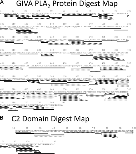
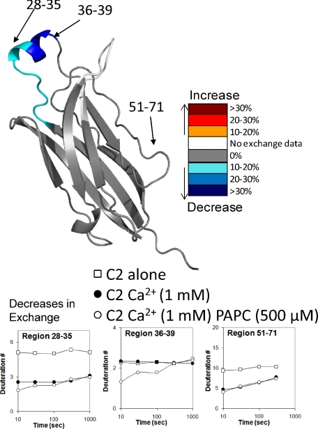
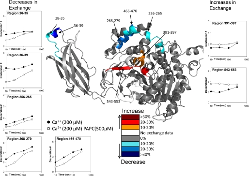
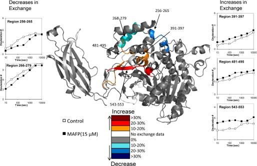
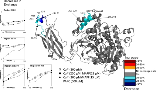
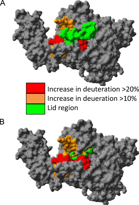
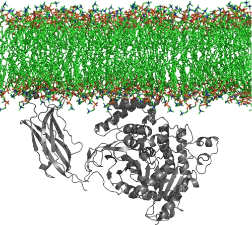
The Group IVA (GIVA) phospholipase A(2) associates with natural membranes in response to an increase in intracellular Ca(2+) along with increases in certain lipid mediators. This enzyme associates with the membrane surface as well as binding a single phospholipid molecule in the active site for catalysis. Employing deuterium exchange mass spectrometry, we have identified the regions of the protein binding the lipid surface and conformational changes upon a single phospholipid binding in the absence of a lipid surface. Experiments were carried out using natural palmitoyl arachidonyl phosphatidylcholine vesicles with the intact GIVA enzyme as well as the isolated C2 and catalytic domains. Lipid binding produced changes in deuterium exchange in eight different regions of the protein. The regions with decreased exchange included Ca(2+) binding loop one, which has been proposed to penetrate the membrane surface, and a charged patch of residues, which may be important in interacting with the polar head groups of phospholipids. The regions with an increase in exchange are all located either in the hydrophobic core underneath the lid region or near the lid and hinge regions from 403 to 457. Using the GIVA phospholipase A(2) irreversible inhibitor methyl-arachidonyl fluorophosphonate, we were able to isolate structural changes caused only by pseudo-substrate binding. This produced results that were very similar to natural lipid binding in the presence of a lipid interface with the exception of the C2 domain and region 466-470. This implies that most of the changes seen in the catalytic domain are due to a substrate-mediated, not interface-mediated, lid opening, which exposes the active site to water. Finally experiments carried out with inhibitor plus phospholipid vesicles showed decreases at the C2 domain as well as charged residues on the putative membrane binding surface of the catalytic domain revealing the binding sites of the enzyme to the lipid surface.


Figures

FIGURE 1.
FIGURE 1.
Pepsin-digested peptide coverage map of GIVA PLA2 and C2 domain. Identified and analyzed pepsin-digested peptides are shown underneath the primary sequence of GIVA PLA2. Only the peptides in bold lines are used in this study.
FIGURE 2.
FIGURE 2.
Deuterium exchange of the isolated C2 domain binding the phospholipid membrane in the presence of Ca2+. DXMS was performed on the C2 domain under the following three conditions: 0 and 1 mm Ca2+ without PAPC, as well as 1 mm Ca2+ with PAPC. The numbers of incorporated deuterons for three regions are shown: 28-35, 36-39, and 51-71. Changes in deuteration between the 1 mm Ca2+ and 1 mm Ca2+ + 500 μm PAPC conditions greater then 10% are represented in color on the crystal structure (PDB 1RLW) (see legend).
FIGURE 3.
FIGURE 3.
Deuterium exchange of the GIVA PLA2 binding the phospholipid membrane in the presence of Ca2+. Ca2+ was present at 200 μm, and vesicles were present at 500 μm. The numbers of incorporated deuterons at four time points in seven different regions, 28-35, 36-39, 256-265, 268-279, 391-397, 466-470, and 543-553, in GIVA PLA2 are plotted. Decreases or increases in deuteration greater then 10% are represented by the color scheme in the legend.
FIGURE 4.
FIGURE 4.
Deuterium exchange upon binding of 15μm MAFP. The number of incorporated deuterons at seven time points in five different regions, 258-265, 268-279, 391-397, 481-495, and 543-553, in GIVA PLA2 are plotted. Decreases or increases in deuteration greater then 10% are represented by the color scheme in the legend.
FIGURE 5.
FIGURE 5.
Deuterium exchange upon Ca2+-mediated PAPC vesicle binding in the presence of 15μm MAFP. Ca2+ was present at 200 μm, and vesicles were present at 500 μm. The numbers of incorporated deuterons at five time points in four different regions, 28-35, 36-39, 268-279, and 466-470, in GIVA PLA2 are plotted. Possible amino acids that may interact with the lipid surface have been shown in stick form. Decreases or increases in deuteration greater then 10% are represented by the color scheme in the legend.
FIGURE 6.
FIGURE 6.
Opening of the lid induced by inhibitor binding. A, the molecular surface of the enzyme with areas that show an increase in exchange upon inhibitor binding is colored red or orange, and the lid region from 415 to 432 is colored green. B, the lid region surface is removed and shown as a ribbon with most areas in red underneath the lid. The deuterium exchange data imply that the lid region from 415 to 432 shifts position to expose the regions underneath. Molecular surface was generated using the Swiss-PdbViewer.
FIGURE 7.
FIGURE 7.
Hypothetical model of initial membrane binding before lid opening. Ca2+ binding at the C2 domain causes translocation of the enzyme to the surface and penetration of the hydrophobic core of the membrane. There seems to be no penetration of the catalytic domain, and its interactions with the surface are mediated through electrostatic contacts from regions 268-279 and 466-470 to PC head groups. The model was generated using PyMOL.

References

    1. Schaloske, R. H., and Dennis, E. A. (2006) Biochim. Biophys. Acta 1761 1246-1259 - PubMed
    1. Six, D. A., and Dennis, E. A. (2000) Biochim. Biophys. Acta 1488 1-19 - PubMed
    1. Clark, J. D., Lin, L. L., Kriz, R. W., Ramesha, C. S., Sultzman, L. A., Lin, A. Y., Milona, N., and Knopf, J. L. (1991) Cell 65 1043-1051 - PubMed
    1. Kramer, R. M., Roberts, E. F., Manetta, J., and Putnam, J. E. (1991) J. Biol. Chem. 266 5268-5272 - PubMed
    1. Funk, C. D. (2001) Science 294 1871-1875 - PubMed

Publication types

MeSH terms

Substances