. 2008 Oct 31;283(44):30174-83.
doi: 10.1074/jbc.M805318200. Epub 2008 Aug 27.

Expression of S100A6 in cardiac myocytes limits apoptosis induced by tumor necrosis factor-alpha

Affiliations

Expression of S100A6 in cardiac myocytes limits apoptosis induced by tumor necrosis factor-alpha

James N Tsoporis et al. J Biol Chem. .

Abstract

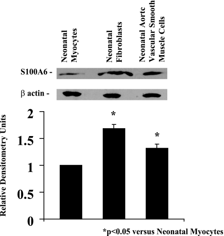
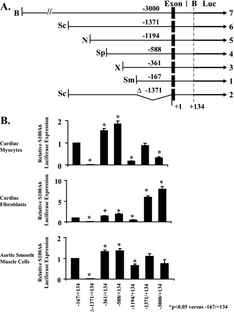
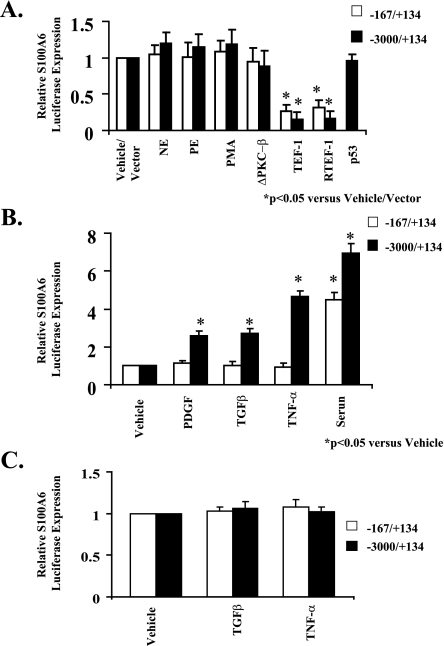
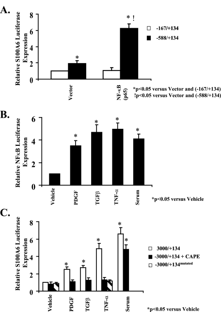
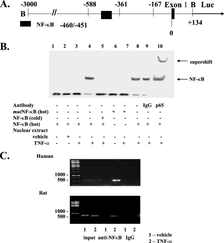
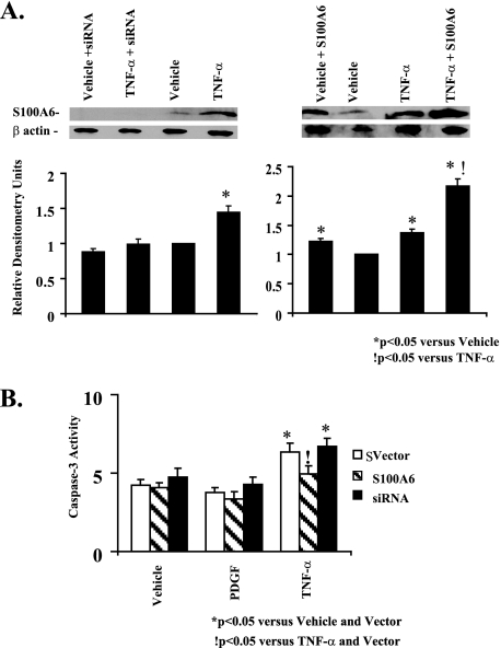
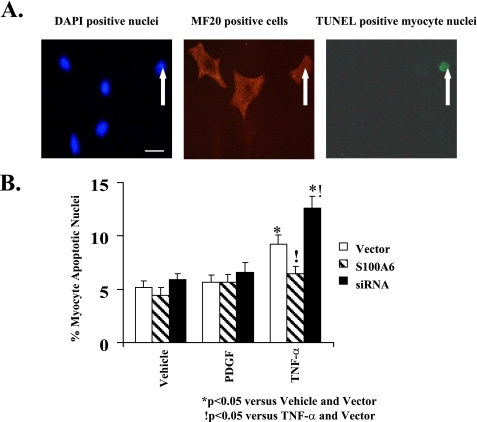
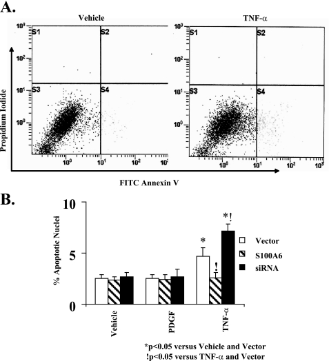
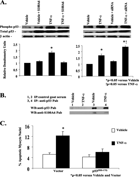
S100A6 is induced in myocardium post-infarction in vivo and in response to growth factors and inflammatory cytokines in vitro. Forced expression of S100A6 in cardiomyocytes inhibits regulation of cardiac specific gene expression in response to trophic stimulation. To define regulation and function of S100A6, we characterized the human S100A6 promoter and mapped upstream regulatory elements in rat neonatal cardiac myocytes, fibroblasts, and vascular smooth muscle cells and defined a functional role for S100A6 in tumor necrosis factor-alpha-induced myocyte apoptosis. The functional S100A6 promoter was localized to region -167/+134 containing 167 upstream base pairs. The S100A6 promoter is regulated by positive (-361/-167 and -588/-361) and negative (-1371/-1194) elements. Tumor necrosis factor-alpha induced the maximal S100A6 promoter and transcription factor NF-kappaB (p65 subunit). Electrophoretic mobility shift showed that tumor necrosis factor-alpha induced p65 binding to a potential NF-kappaB-binding site at -460/-451. Chromatin immunoprecipitation analysis revealed p65 is recruited to the S100A6 promoter upon tumor necrosis factor-alpha stimulation. The NF-kappaB inhibitor caffeic acid phenethyl ester and mutation of the NF-kappaB-binding site inhibited S100A6 promoter activation by tumor necrosis factor-alpha. Tumor necrosis factor-alpha induced cardiac myocyte apoptosis. Specific inhibition of S100A6 using a small interfering RNA directed against S100A6 potentiated tumor necrosis factor-alpha-induced myocyte apoptosis, whereas overexpression of S100A6 by gene transfer prevented tumor necrosis factor-alpha-induced myocyte apoptosis by interfering with p53 phosphorylation. These results demonstrate that S100A6 is induced by tumor necrosis factor-alpha via an NF-kappaB-dependent mechanism, serving a role in homeostasis to limit tumor necrosis factor-alpha-induced apoptosis by regulating p53 phosphorylation.


Figures

FIGURE 1.
FIGURE 1.
Western blot analysis of S100A6 and β-actin proteins in rat neonatal cultured myocytes, fibroblasts, and aortic smooth muscle cells. Protein extracts from each treatment group were analyzed by Western blotting using goat anti-S100A6 and mouse anti-β-actin antibodies. Respective relative densitometry units are displayed below blots. *, p < 0.05 versus neonatal myocytes. n = 3.
FIGURE 2.
FIGURE 2.
Human S100A6 promoter-luciferase reporter constructs. A, using the full-length pBS construct, shorter fragments of the human S100A6 gene (solid boxes were prepared by restriction digests to generate deletion constructs). Restriction enzymes are abbreviated as follows: B, BamHI; Sc, ScaI; N, NcoI; Sp, SphI; X, XhoI; Sm, SmaI. The black arrows (luciferase (Luc)) represent the coding region of firefly luciferase. B, neonatal rat cardiac myocytes, cardiac fibroblasts, and aortic smooth muscle cells were co-transfected with the appropriate S100A6 promoter-luciferase construct and RSV-CAT. The bars shown are the mean ± S.E. of luciferase expression (normalized to the RSV-CAT activity) relative to pGBS-167 in the same cell type, which has been assigned the value of 1. *, p < 0.05 relative to pGBS-167. n = 6.
FIGURE 3.
FIGURE 3.
Selective stimulation of the S100A6 promoter by trophic stimuli. A, myocyte cultures were co-transfected with the human S100A6 promoter-luciferase reporter constructs and treated for 48 h with either norepinephrine (NE, 20 μm), phenylephrine (PE, 20 μm), PMA (10 nm), or co-transfected with the expression plasmids ΔPKC-β, TEF-1, RTEF-1, or p53. B, myocyte cultures were co-transfected with the human S100A6 promoter-luciferase reporter constructs and treated for 48 h with either PDGF (5 ng/ml), TGFβ (5 ng/ml), TNF-α (5 ng/ml), or 5% serum. C, non-myocyte (fibroblast) cultures were co-transfected with the human S100A6 promoter-luciferase reporter constructs and treated for 48 h with either TGFβ (5 ng/ml) or TNF-α (5 ng/ml). Bars shown are mean ± S.E. of luciferase expression (normalized to the RSV-CAT activity) relative to vehicle treatment of each deletion fragment (assigned the value of 1). *, p < 0.05 versus vehicle/vector. n = 6.
FIGURE 4.
FIGURE 4.
NF-κB activation is required for TNF-α induction of the human S100A6 promoter. A, myocyte cultures were co-transfected with either the -167/+134 or -588/+134 human S100A6 promoter-luciferase reporter constructs in combination with the p65 expression plasmid or empty vector as indicated. B, myocyte cultures were co-transfected with the human NF-κB promoter-luciferase reporter constructs and treated for 48 h with either PDGF (5 ng/ml), TGFβ (5 ng/ml), TNF-α (5 ng/ml), 5% serum, or vehicle. C, myocyte cultures were co-transfected with either the -3000/+134 or -3000/+134mutated (containing point mutations of the NF-κB-binding site) human S100A6 promoter-luciferase reporter constructs as indicated, and the -3000/+134 transfected cells were pretreated with the NF-κB inhibitor CAPE (50 μg/ml) or vehicle for 2 h and then treated for 48 h with either PDGF (5 ng/ml), TGFβ (5 ng/ml), TNF-α (5 ng/ml), or 5% serum. Bars shown are mean ± S.E. of luciferase expression (normalized to the RSV-CAT activity) relative to -167/+134 with vector alone (A) or vehicle control (B and C) (assigned the value of 1). *, p < 0.05 versus vehicle or vector.!, p < 0.05 versus -588/+134 and vector alone. n = 6.
FIGURE 5.
FIGURE 5.
NF-κB binds to its putative site in the S100A6 promoter in vitro and in vivo in response to TNF-α. A, schematic representation of the -3000/+134 S100A6 promoter fragment containing the first exon. The location of the restriction sites (B, BamHI) used to generate the fragment and location of the NF-κB site are shown. B, electromobility shift assay using nuclear extracts from myocyte cultures treated with TNF-α (5 ng/ml) or vehicle as indicated. The binding of NF-κB was confirmed by point mutations (mut) in NF-κB oligonucleotides, addition of 100-fold excess cold competitor oligonucleotides, and by supershift with an antibody to NF-κB p65 as indicated. C, myocyte cultures were co-transfected with the -588/+134 human S100A6 promoter-luciferase reporter construct as indicated and treated with TNF-α (5 ng/ml) or vehicle for 1 h then harvested and then subjected to chromatin immunoprecipitation analysis using NF-κB p65 antibody or a nonspecific anti-rabbit IgG. The isolated DNA fragments were amplified by PCR using both human (C, top) and rat (C, bottom) S100A6-specific primers. Input, nonprecipitated chromatin.
FIGURE 6.
FIGURE 6.
TNF-α induction of S100A6 in cardiac myocytes. A, representative Western blots of S100A6 andβ-actin in myocyte cultures transfected with either vector alone or the human S100A6 expression plasmid or an siRNA duplex against S100A6 and treated for 48 h with either vehicle or TNF-α (5 ng/ml). Respective densitometry units relative to vehicle control are displayed below blots. *, p < 0.05 versus vehicle and vector.!, p < 0.05 versus TNF-α and vector. n = 3. B, myocyte cultures were transfected with either a human S100A6 expression plasmid or an siRNA duplex against S100A6 or empty vector and treated for 48 h with either PDGF (5 ng/ml), TNF-α (5 ng/ml), or vehicle. Myocytes were assessed for caspase-3 activity by the EnzChek caspase-3 assay kit. The results are the mean ± S.E. of 6–8 different experiments. *, p < 0.05 versus vehicle and/or vector.!, p < 0.05 versus TNF-α and vector alone.
FIGURE 7.
FIGURE 7.
S100A6 modulation of TNF-α-induced myocyte apoptosis as assessed by TUNEL staining. A, representative photomicrographs (×400) of 4′,6-diamidino-2-phenylindole (DAPI)-nuclei (left) (bar = 10 μm), MF20-stained cardiac myocytes (middle), and TUNEL-positive (right) cardiac myocyte(s) treated with TNF-α (5 ng/ml). White arrow indicates myocyte apoptotic nucleus as visualized by TUNEL staining. B, myocyte cultures were transfected with either an S100A6 expression plasmid or an siRNA duplex against S100A6 and treated for 48 h with either PDGF (5 ng/ml), TNF-α (5 ng/ml), or vehicle. TUNEL-positive myocyte nuclei were counted and expressed as percentage of total myocyte nuclei. The results are the mean ± S.E. from random fields in blinded experiments. A minimum of 10 high power fields were scored per experiment of six different experiments. *, p < 0.05 versus vehicle/vector.!, p < 0.05 versus TNF-α/vector.
FIGURE 8.
FIGURE 8.
S100A6 modulation of TNF-α-induced myocyte apoptosis as defined by flow cytometry. A, representative graphs showing the proportion of apoptotic myocytes (FITC-annexin V positive/propidium iodide negative) indicated in the lower right quadrant following treatment with either vehicle (left graph) or TNF-α (5 ng/ml) (right graph). B, myocyte cultures were transfected with either a human S100A6 expression plasmid or an siRNA duplex against S100A6 or empty vector and treated for 48 h with either PDGF (5 ng/ml), TNF-α (5 ng/ml), or vehicle. Myocytes were assessed for apoptosis by FITC annexin V/propidium iodide staining and flow cytometry. The results are the mean ± S.E. from 6 to 8 different experiments. *, p < 0.05 versus vehicle/vector. !, p < 0.05 versus TNF-α and vector.
FIGURE 9.
FIGURE 9.
S100A6 modulation of TNF-α-induced myocyte apoptosis requires p53. A, representative Western blot of phospho-p53, total p53, and β-actin in myocyte cultures transfected with either vector alone, human S100A6 expression plasmid, or an siRNA duplex against S100A6 and treated for 48 h with either vehicle or TNF-α (5 ng/ml). Respective relative densitometry units are displayed below blots. *, p < 0.05 versus vehicle and vector.!, p < 0.05 versus TNF-α and vector. n = 3. B, co-immunoprecipitation of p53 and S100A6 in TNF-α-treated myocyte cultures. Aliquots of lysates of myocyte cultures treated with either vehicle or TNF-α were incubated with control goat serum (lanes 1 and 2) or anti-p53 polyclonal antibody (lanes 3 and 4), followed by incubation with protein A-Sepharose. The immune complexes were dissociated and analyzed by Western blotting with anti-S100A6 or anti-p53 antibodies as indicated in each blot. C, myocyte cultures were co-transfected with a mutant p53His175 plasmid acting as a dominant negative and treated for 48 h with either TNF-α (5 ng/ml), 5% serum, or vehicle. The results are the mean ± S.E. of six different experiments. *, p < 0.05 versus vehicle/vector.

References

    1. Kligman, D., and Hilt, D. C. (1988) Trends Biochem. Sci. 13 437-443 - PubMed
    1. Zimmer, D. B., Cornwall, E. H., Landar, A., and Song, W. (1995) Brain Res. Bull. 37 417-429 - PubMed
    1. Schafer, B. W., and Heizmann, C. W. (1996) Trends Biochem. Sci. 21 134-140 - PubMed
    1. Kuznicki, J., Filipek, A., Heimann, P., Kaczmarek, L., and Kaminska, B. (1989) FEBS Lett. 254 141-144 - PubMed
    1. Engelkamp, D., Schafer, B. W., Erne, P., and Heizmann, C. W. (1992) Biochemistry 31 10258-10264 - PubMed

Publication types