. 2008 Nov;82(22):11318-30.
doi: 10.1128/JVI.01052-08. Epub 2008 Aug 27.

The M, E, and N structural proteins of the severe acute respiratory syndrome coronavirus are required for efficient assembly, trafficking, and release of virus-like particles

Affiliations

The M, E, and N structural proteins of the severe acute respiratory syndrome coronavirus are required for efficient assembly, trafficking, and release of virus-like particles

Y L Siu et al. J Virol. 2008 Nov.

Abstract

The production of virus-like particles (VLPs) constitutes a relevant and safe model to study molecular determinants of virion egress. The minimal requirement for the assembly of VLPs for the coronavirus responsible for severe acute respiratory syndrome in humans (SARS-CoV) is still controversial. Recent studies have shown that SARS-CoV VLP formation depends on either M and E proteins or M and N proteins. Here we show that both E and N proteins must be coexpressed with M protein for the efficient production and release of VLPs by transfected Vero E6 cells. This suggests that the mechanism of SARS-CoV assembly differs from that of other studied coronaviruses, which only require M and E proteins for VLP formation. When coexpressed, the native envelope trimeric S glycoprotein is incorporated onto VLPs. Interestingly, when a fluorescent protein tag is added to the C-terminal end of N or S protein, but not M protein, the chimeric viral proteins can be assembled within VLPs and allow visualization of VLP production and trafficking in living cells by state-of-the-art imaging technologies. Fluorescent VLPs will be used further to investigate the role of cellular machineries during SARS-CoV egress.


Figures

FIG. 1.
FIG. 1.
Production of SARS-CoV VLPs by transfected Vero E6 cells. (A) Coexpression of M, E, and N is necessary for efficient secretion of SARS-CoV VLPs by Vero E6 cells at 24 and 48 h posttransfection. Monolayers of Vero E6 cells were transfected with plasmids driving the expression of the SARS-CoV structural proteins M, E, and Flag-tagged N as specified at the top of each lane. Protein expression in cell lysates and in VLPs isolated from culture medium was analyzed by Western blotting at 24 and 48 h posttransfection, as indicated below the corresponding panels. Samples were heat denatured and reduced with dithiothreitol before loading. The M and E proteins were detected with rabbit polyclonal antibodies produced against the C-terminal extremity of each proteins. The N protein was detected with the M2 monoclonal antibody recognizing the Flag tag. Blots were exposed for 1 min for signal detection except for the detection of E contained in pellets from ultracentrifuged culture medium (bottom panels), for which blots were exposed for 10 min. The molecular mass (in kilodaltons) and the migration of protein standards are shown between the blots. (B) Use of the bicistronic pIRES-M-E vector restrains the E expression level and favors the production of M-E-N VLPs. Vero E6 cells were transfected with the indicated plasmid combinations, and the cell lysates and medium were analyzed at 48 h posttransfection as in panel A. To ensure better detection of E, VLPs were concentrated four times more than in panel A. Blots were exposed for 10 s for signal detection except for the detection of E contained in pellets from ultracentrifuged culture medium (right bottom panel), for which the blot was exposed for 1 min. (C) Secreted viral structural proteins cosediment in sucrose gradient. Three 75-cm2 dishes of Vero E6 cells were transfected with plasmids driving the expression of SARS-CoV structural proteins M, E, and Flag-tagged N either individually or in combination. Protein expression in cell lysates and in pellets from culture medium ultracentrifuged on 20% sucrose cushion was controlled by Western blot at 48 h posttransfection (left panel). Resuspended pellets from ultracentrifuged cell medium were then loaded on a 20 to 60% discontinuous sucrose gradient and subjected to fractionation by ultracentrifugation. Twenty fractions of 600 μl were collected (1 to 20, from lightest to heaviest). The nature of viral proteins associated with each fraction was determined by Western blotting (a to e). Portions (15 μl) of samples were heat denatured and reduced with dithiothreitol before loading. The molecular mass (in kilodaltons) and the migration of protein standards are shown on the right sides of the blots. Samples from (i) pIRES-M, pIRES-M plus pCDNA-E, and pIRES-M plus pCDNA-E plus pcDNA-Nflag and (ii) pcDNA-E and pIRES-M plus pcDNA-Nflag were generated in two separate experiments. Ctrl, nontransfected cells; E, pCDNA-E; M, pIRES-M; M-E, pIRES-M-E; M + Nflag, pIRES-M plus pcDNA-Nflag; M-E + Nflag, pIRES-M-E plus pcDNA-Nflag; M + E, pIRES-M + pcDNA-E; M + E + Nflag, pIRES-M plus pcDNA-E plus pcDNA-Nflag.
FIG. 2.
FIG. 2.
S-glycoprotein trimers are incorporated onto SARS-CoV M, E, and N-containing VLPs. pcDNA-S plasmid was cotransfected with pIRES-M-E and pcDNA-Nflag vectors. The culture medium was harvested at 48 h posttransfection and ultracentrifuged on 20% sucrose cushion, and the pellets were resuspended in TNE buffer and ultracentrifuged on a 20 to 60% discontinuous sucrose gradient. Twenty fractions were collected (1 to 20, from lightest to heaviest) and analyzed by Western blotting. Samples were either heat denatured and reduced with dithiothreitol before loading for detection of Nflag, M, and E or not heated and not reduced for analysis of S. Blots were exposed for 10 s for signal detection, except for E, for which the blots were exposed for 10 min. The highest levels of S, M, N, and E structural proteins were found in fractions 9 and 10 corresponding to 40% sucrose. S protein was detected with mouse polyclonal antibodies raised against purified S trimers. Arrows indicate bands that correspond to trimeric and monomeric forms of S. The molecular mass (in kilodaltons) and the migration of protein standards are shown on the right sides of the blots.
FIG. 3.
FIG. 3.
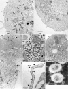
Structural analysis and intracellular distribution of SARS-CoV VLPs. Vero E6 cells were cotransfected with pIRES-M-E, pcDNA-Nflag, and pcDNA-S. At 24 and 48 h posttransfection the cells were fixed, and ultrathin sections were analyzed by electron microscopy (A to G). (A) VLPs were found in intracellular vacuoles (vc) and in vesicles (vs) scattered in the cytosol and bound to the plasma membrane (pm). The arrow points to VLPs attached to the cell surface. (B) Large amount of VLPs within the lumen of the endoplasmic reticulum (er) and within a cytoplasmic vesicle. n, nucleus. (C) Presence of VLPs within vacuoles and vesicles. Arrows point to small VLPs-containing vesicles beneath the plasma membrane. (D) Magnification of a VLP-containing vacuole. Black arrows point to budding events. (E) Compacted VLP-containing vesicles were found beneath the plasma membrane. (F) VLPs bound to the surface of a producer cell. Two membrane-bound VLPs are indicated by arrows. (G) VLPs bound to a cell filopodia. Spikelike protuberances were visible on the VLP surface (arrows). (H) Electron microscopy images of negatively stained VLPs purified from cell medium at 48 h posttransfection. A scale bar is indicated for each picture. Panels A to D and panels F to G correspond to cells fixed at 48 h posttransfection. The image in panel E was taken from cells fixed at 24 h posttransfection. n, nucleus; er, ER; vc, vacuole; vs, vesicle; pm, plasma membrane.
FIG. 4.
FIG. 4.
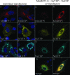
Expression and subcellular distribution of viral structural proteins tagged with fluorescent proteins. Vero E6 cells grown on glass coverslips were either transfected with single plasmids (left panels) or cotransfected (right panels) with the three plasmids encoding the MmRFP1, E, SeYFP, and NeCFP proteins. At 24 h posttransfection, cells were processed for nuclear staining with DAPI dye, fixed, and analyzed under a fluorescence microscope equipped with an ApoTome device to acquire images of optical sections. a, b, g, and h, MmRFP1; c, d, i, and j, SeYFP; e, f, k, and l, NeCFP; m and n, merged images. For all conditions, two representative images are shown side by side.
FIG. 5.
FIG. 5.
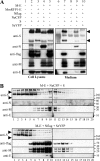
Production of fluorescent VLPs by transfected Vero E6 cells. (A) Determination of optimal plasmid combinations for production of SARS-CoV fluorescent VLPs. Viral proteins from cell lysates (left panel) and sedimented VLPs from medium (right panel) were analyzed by Western blotting. VLPs could be produced when either N or S (lanes 9 and 10) but not M (lane 8) were tagged with fluorescent proteins. (B) Production of fluorescent VLPs and efficiency of incorporation of NeCFP and SeYFP fusion proteins into VLPs. Cells were cotransfected with the specified plasmid combinations (corresponding to lanes 9 and 10 of panel A), and purified VLPs from medium were analyzed by using a sucrose gradient. Tagging S (lower blot) resulted in a greater yield of VLP production compared to tagged N (upper blot). However, the eYFP tag greatly reduced S incorporation into VLPs (lower blot). Arrowheads indicate spike trimers and monomers.
FIG. 6.
FIG. 6.
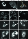
Tracking of fluorescent SARS-CoV VLPs in living cells. (A) Wide-field fluorescence microscopy images showing the accumulation of fluorescent VLPs at the plasma membrane of pIRES-M-E plus pcDNA-NeCFP cotransfected cells (panel b), whereas a strong perinuclear staining was observed in Vero E6 cells expressing NeCFP alone (panel a). (B) Confocal microscopy of living cells expressing M-E-NeGFP VLPs. Four categories of fluorescent signals were observed: a bright and static large perinuclear compartment (white encircling lines), smaller and dimmer actively trafficking vesicles (orange encircling lines), bright dots accumulating at the cell cortex (yellow encircling lines), and dots in the medium surrounding transfected cells (yellow dots). Videos are available in the supplemental material. (C) Treatment of transfected cells with BFA alters the trafficking of fluorescent vesicles. Vero E6 cells were transfected with pIRES-M-E plus pcDNA-NeGFP plasmids. Cells were either not treated (panel a) or treated with 6 mg of BFA/ml for either 4 h (panel d) or overnight (panel g). BFA was then added to untreated cells, and time-lapse acquisitions were performed. Panels b and c show the same cells as in panel a but after 25- and 70-min incubations with the drug. Alternatively, BFA was washed out and recovery after BFA treatment was analyzed (panels e and f and panels h and i). Panels a, b, and c, panels d and e, and panels g and h show the same cells at different time points. Videos illustrating panels b, f, h, and i are provided in the supplemental material.

References

    1. Baudoux, P., C. Carrat, L. Besnardeau, B. Charley, and H. Laude. 1998. Coronavirus pseudoparticles formed with recombinant M and E proteins induce alpha interferon synthesis by leukocytes. J. Virol. 728636-8643. - PMC - PubMed
    1. Bos, E. C., W. Luytjes, H. V. van der Meulen, H. K. Koerten, and W. J. Spaan. 1996. The production of recombinant infectious DI-particles of a murine coronavirus in the absence of helper virus. Virology 21852-60. - PMC - PubMed
    1. Bosch, B. J., C. A. de Haan, and P. J. Rottier. 2004. Coronavirus spike glycoprotein, extended at the carboxy terminus with green fluorescent protein, is assembly competent. J. Virol. 787369-7378. - PMC - PubMed
    1. Bosch, B. J., C. A. de Haan, S. L. Smits, and P. J. Rottier. 2005. Spike protein assembly into the coronavirion: exploring the limits of its sequence requirements. Virology 334306-318. - PMC - PubMed
    1. Campbell, R. E., O. Tour, A. E. Palmer, P. A. Steinbach, G. S. Baird, D. A. Zacharias, and R. Y. Tsien. 2002. A monomeric red fluorescent protein. Proc. Natl. Acad. Sci. USA 997877-7882. - PMC - PubMed

Publication types

MeSH terms