. 2008 Nov;82(21):10613-24.
doi: 10.1128/JVI.01241-08. Epub 2008 Aug 27.

Herpes simplex virus gE/gI and US9 proteins promote transport of both capsids and virion glycoproteins in neuronal axons

Affiliations

Herpes simplex virus gE/gI and US9 proteins promote transport of both capsids and virion glycoproteins in neuronal axons

Aleksandra Snyder et al. J Virol. 2008 Nov.

Abstract

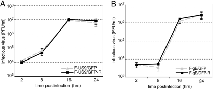
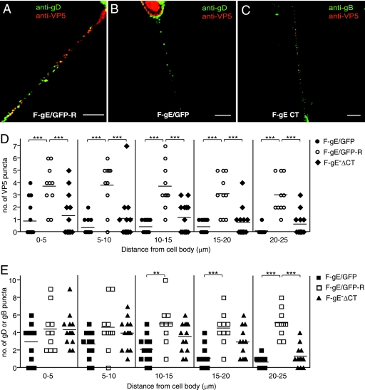
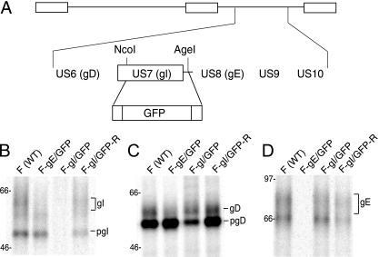
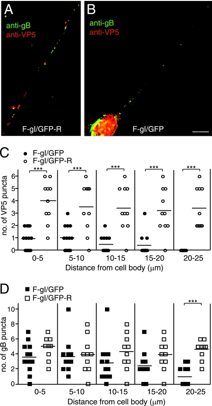
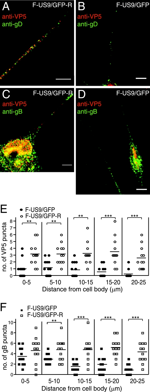
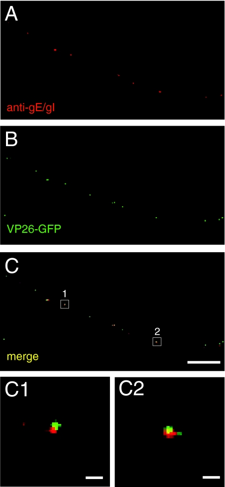
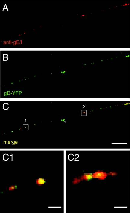
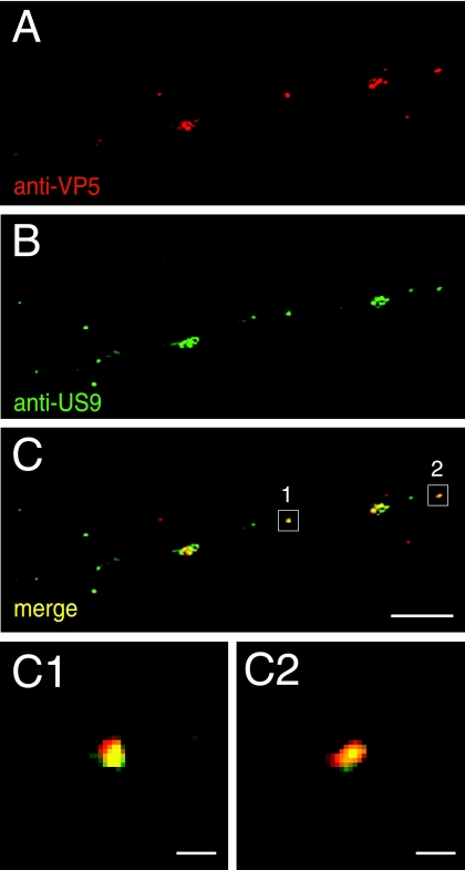
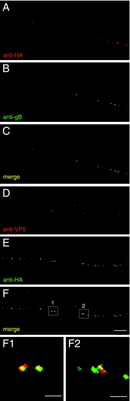
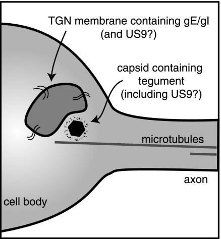
Following reactivation from latency, alphaherpesviruses replicate in sensory neurons and assemble capsids that are transported in the anterograde direction toward axon termini for spread to epithelial tissues. Two models currently describe this transport. The Separate model suggests that capsids are transported in axons independently from viral envelope glycoproteins. The Married model holds that fully assembled enveloped virions are transported in axons. The herpes simplex virus (HSV) membrane glycoprotein heterodimer gE/gI and the US9 protein are important for virus anterograde spread in the nervous systems of animal models. It was not clear whether gE/gI and US9 contribute to the axonal transport of HSV capsids, the transport of membrane proteins, or both. Here, we report that the efficient axonal transport of HSV requires both gE/gI and US9. The transport of both capsids and glycoproteins was dramatically reduced, especially in more distal regions of axons, with gE(-), gI(-), and US9-null mutants. An HSV mutant lacking just the gE cytoplasmic (CT) domain displayed an intermediate reduction in capsid and glycoprotein transport. We concluded that HSV gE/gI and US9 promote the separate transport of both capsids and glycoproteins. gE/gI was transported in association with other HSV glycoproteins, gB and gD, but not with capsids. In contrast, US9 colocalized with capsids and not with membrane glycoproteins. Our observations suggest that gE/gI and US9 function in the neuron cell body to promote the loading of capsids and glycoprotein-containing vesicles onto microtubule motors that ferry HSV structural components toward axon tips.


Figures

FIG. 1.
FIG. 1.
Replication of HSV US9 and gE mutants in human neurons. Differentiated SK-N-SH neurons were infected using 5 PFU/cell with either F-US9/GFP (US9) or F-US9/GFP-R (a repaired version of F-US9/GFP) (A) or with F-gE/GFP (gE) or F-gE/GFP-R (a repaired version of F-gE/GFP) (B). At various times, triplicate wells of cells and cell culture supernatants were harvested and sonicated, and the titer of infectious virus was determined using Vero cells. Standard deviations are shown as error bars.
FIG. 2.
FIG. 2.
Axonal transport of capsids and glycoproteins in human neurons infected with HSV gE mutants. SK-N-SH neurons were infected with the repaired F-gE/GFP-R (A), the gE-null mutant F-gE/GFP (B), or a gE mutant lacking the gE CT domain, F-gEΔCT (C), for 18 h; fixed; permeabilized; and stained with mouse anti-VP5 MAb and rabbit anti-gD polyclonal antibodies (A and B) or mouse anti-VP5 MAb and rabbit anti-gB polyclonal antibodies (C), followed by Texas red-conjugated donkey anti-mouse and Cy-5-conjugated anti-rabbit secondary antibodies. Scale bars, 5 μm. Shown is the quantification of VP5 puncta (D) and gB or gD puncta (E) in segments of neuronal axons (0 to 5, 5 to 10, 10 to 15, 15 to 20, or 20 to 25 um) measured from the neuronal cell body. Puncta numbers were obtained from 13 F-gE/GFP-infected, 10 F-gE/GFP-R-infected, and 13 FgE ΔCT-infected neurons involving four independent experiments. Each symbol represents the number of VP5 or glycoprotein puncta observed in the indicated axon segment. Statistically significant values are shown as asterisks, with a P value of <0.05 marked as two asterisks and a P value of <0.001 marked as three asterisks.
FIG. 3.
FIG. 3.
Construction and characterization of an HSV gI mutant. (A) Schematic of the HSV unique short component, including the US6 to US10 genes. In F-gI/GFP, the gI (US7) coding sequences between NcoI and AgeI restriction sites were replaced with GFP sequences. A second virus, in which gI sequences were repaired, was produced by cotransfecting Vero cells with F-gI/GFP DNA and a plasmid containing the gE and gI genes. (B to D) R970 cells were infected with either wild-type HSV (WT), F- gE/GFP, F-gI/GFP, or F-gI/GFP-R using 10 PFU/cell for 6 h, and the cells were labeled with [35S]methionine/cysteine for 3 h. gI (B), gD (C), and gE (D) were immunoprecipitated from detergent extracts of the cells using MAb 3104, 3114, or DL6, respectively, and gel electrophoresis was performed. The positions of mature glycoproteins, gI, gD, and gE, and immature forms of glycoproteins, pgI and pgD, and marker proteins of 97, 66, and 46 kDa are indicated.
FIG. 4.
FIG. 4.
Axonal transport of capsids and glycoproteins in human neurons infected with the HSV gI mutant. SK-N-SH neurons were infected with the repaired F-gI/GFP-R (A) or the gI mutant F-gI/GFP (B) for 18 h, fixed, permeabilized, and stained with mouse anti-VP5 MAb and rabbit anti-gB polyclonal antibodies, followed by Texas red-conjugated donkey anti-mouse and Cy-5-conjugated anti-rabbit secondary antibodies. Scale bars, 5 μm. (C and D) Quantification of VP5 puncta (C) and gB puncta (D) in different segments of neuronal axons measured from the neuronal-cell body. VP5 or gB puncta were counted in 13 F-gI/GFP-infected and 10 F-gI/GFP-R-infected neurons involving four separate experiments. Each symbol represents the number of puncta present in an individual axonal segment. Statistically significant values are shown as asterisks, with P < 0.05 marked as two asterisks and P < 0.001 marked as three asterisks.
FIG. 5.
FIG. 5.
Axonal transport of capsids and membrane glycoproteins in human neurons infected with an HSV US9 mutant. Differentiated SK-N-SH neurons were infected with either the repaired F-US9/GFP-R (A and C) or the US9-null mutant F-US9/GFP (B and D) for 18 h, fixed, permeabilized, and stained with mouse anti-VP5 MAb and rabbit anti-gD polyclonal antibodies (A and B) or mouse anti-VP5 MAb and rabbit anti-gB polyclonal antibodies (C and D), followed by Texas red-conjugated donkey anti-mouse and Cy-5-conjugated anti-rabbit secondary antibodies. Scale bars, 5 μm. (E and F) Quantification of VP5 puncta (E) and gB puncta (F) in axonal segments measured from the neuronal-cell body. Puncta were counted in the axons of 13 F-US9/GFP-infected and 13 F-US9/GFP-R-infected neurons involving four independent experiments. Each symbol represents the number of puncta present in an individual axonal segment. Statistically significant values are shown as asterisks, with a P value of <0.05 marked as two asterisks and a P value of <0.001 marked as three asterisks.
FIG. 6.
FIG. 6.
gE does not associate with capsids in axons of HSV-infected neurons. SK-N-SH neurons were infected with F-VP26-GFP, which expresses a fluorescent capsid protein for 24 h, and then the neurons were fixed, permeabilized, and stained with rat anti-gE/gI polyclonal antibodies, followed by Texas red-conjugated donkey anti-mouse secondary antibodies. Representative results from four independent experiments are shown. (A) Rat anti-gE/gI staining. (B) VP26-GFP fusion protein fluorescence. (C) Merged fluorescence. (C1 and C2) Higher magnifications of the areas boxed in panel C. Scale bars, 5 μm (A to C) and 1 μm (C1 and C2).
FIG. 7.
FIG. 7.
gE/gI associates with glycoprotein gD in axons of HSV-infected neurons. SK-N-SH neurons were infected with SC16 gD-YFP for 24 h, fixed, permeabilized, and stained with rat anti-gE/gI polyclonal antibodies, followed by Texas red-conjugated donkey anti-mouse secondary antibodies. Representative panels of four independent experiments are shown. (A) Rat anti-gE/gI staining. (B) SC16-gD-YFP fusion protein fluorescence. (C) Merged fluorescence. (C1 and C2) Higher magnifications of the boxed areas in panel C. Scale bars, 5 μm (A to C) and 1 μm (C1 and C2). The star indicates gD-YFP fluorescence associated with a particle that was not present within the axon shown.
FIG. 8.
FIG. 8.
US9-specific antibodies stain capsid puncta in axons of HSV-infected neurons. SK-N-SH neurons were infected with wild-type HSV for 18 h, fixed, permeabilized, and stained with mouse anti-VP5 MAb and simultaneously with affinity-purified rabbit anti-US9 antibodies, followed by Texas red-conjugated donkey anti-mouse secondary antibodies and fluorescein isothiocyanate-conjugated donkey anti-rabbit secondary antibodies. Panels A to C are representative of three independent experiments. (A) Mouse anti-VP5 staining. (B) Rabbit anti-US9 staining. (C) Merged fluorescence. (C1 and C2) Higher magnifications of the areas boxed in panel C. Scale bars, 5 μm (A to C) and 1 μm (C1 and C2).
FIG. 9.
FIG. 9.
HA epitope-tagged US9 closely associates with capsids in neuronal axons. SK-N-SH neurons were infected with F-US9-HA for 18 h, fixed, permeabilized, and stained with mouse anti-HA MAb and rabbit anti-gB polyclonal antibodies (A to C) or rabbit anti-HA polyclonal antibodies and mouse anti-VP5 MAb (D to F). Secondary antibodies were Texas red-conjugated donkey anti-mouse secondary antibodies and fluorescein isothiocyanate-conjugated donkey anti-rabbit. The images are representative of three independent experiments. (F1 and F2) High magnifications of axonal segments 1 and 2 in panel F. Scale bars, 5 μm (A to F) and 1 μm (F1 and F2).
FIG. 10.
FIG. 10.
Model showing how gE/gI and US9 might facilitate the loading of HSV capsids onto microtubules within neuronal-cell bodies. TGN membrane vesicles containing gE/gI and present in neuronal-cell bodies are adjacent to microtubules and serve as platforms for the assembly or loading of HSV tegument-coated capsids onto microtubule motors prior to transport into and down axons. HSV US9 may be a membrane protein and act similarly, or it may be a tegument protein bound to the surfaces of capsids.

Similar articles

Cited by

References

    1. Antinone, S. E., and G. A. Smith. 2006. Two modes of herpesvirus trafficking in neurons: membrane acquisition directs motion. J. Virol. 8011235-11240. - PMC - PubMed
    1. Brideau, A. D., B. W. Banfield, and L. W. Enquist. 1998. The Us9 gene product of pseudorabies virus, an alphaherpesvirus, is a phosphorylated, tail-anchored type II membrane protein. J. Virol. 724560-4570. - PMC - PubMed
    1. Brideau, A. D., J. P. Card, and L. W. Enquist. 2000. Role of pseudorabies virus Us9, a type II membrane protein, in infection of tissue culture cells and the rat nervous system. J. Virol. 74834-845. - PMC - PubMed
    1. Brideau, A. D., M. G. Eldridge, and L. W. Enquist. 2000. Directional transneuronal infection by pseudorabies virus is dependent on an acidic internalization motif in the Us9 cytoplasmic tail. J. Virol. 744549-4561. - PMC - PubMed
    1. Brown, A. 2003. Axonal transport of membranous and nonmembranous cargoes: a unified perspective. J. Cell Biol. 160817-821. - PMC - PubMed

Publication types

Substances

LinkOut - more resources