

Interleukin-18 binding protein transgenic mice are protected against ischemic acute kidney injury
- PMID: 18753296
- PMCID: PMC2584896
- DOI: 10.1152/ajprenal.90288.2008
Interleukin-18 binding protein transgenic mice are protected against ischemic acute kidney injury
Abstract
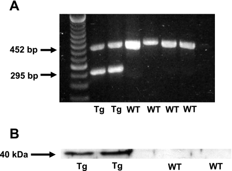
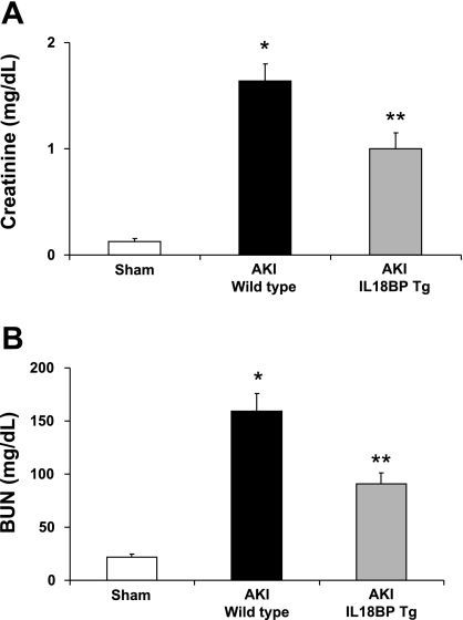
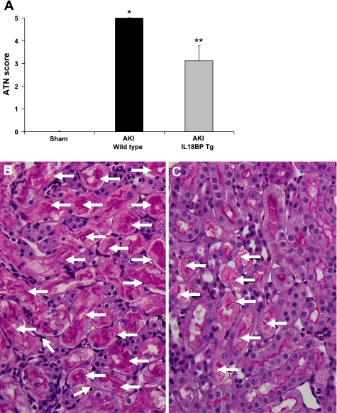
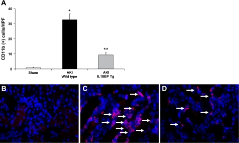
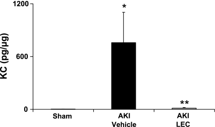
IL-18 function is neutralized in IL-18 binding protein transgenic (IL-18BP Tg) mice. First, we determined whether IL-18BP Tg mice are protected against ischemic acute kidney injury (AKI). Ischemic AKI was induced by bilateral renal pedicle clamping. IL-18BP Tg mice were functionally and histologically protected against ischemic AKI as determined by blood urea nitrogen, serum creatinine, and acute tubular necrosis score. We have demonstrated that the injurious effect of IL-18 in the kidney is independent of neutrophils and lymphocytes. Thus the effect of IL-18 inhibition on renal macrophage infiltration was determined. The number of macrophages was significantly reduced in IL-18BP Tg compared with wild-type kidneys. To determine the cytokines and chemokines that are dependent on IL-18, we performed flow cytometry based assays. Multiple chemokines/cytokines, IL-3, IL-6, IL-15, IL-18, leukemia inhibitory factor, macrophage colony-stimulating factor, macrophage inflammatory protein-2, granulocyte-macrophage colony-stimulating factor, and monocyte chemotactic protein-1 were significantly increased in AKI vs. sham kidneys. Only CXCL1 (also known as KC or IL-8) was significantly increased in AKI vs. sham kidneys and significantly reduced in IL-18BP Tg AKI vs. wild-type AKI kidneys. To determine whether macrophages are the source of CXCL1 in the kidney, we depleted macrophages with liposomal encapsulated clodronate. CXCL1 was significantly decreased in macrophage-depleted vs. control AKI mice. In summary, in ischemic AKI in mice, 1) IL-18BP Tg mice are functionally and histologically protected, 2) macrophage infiltration in the kidney and CXCL1 are significantly reduced in IL-18BP Tg mice, and 3) macrophage depletion significantly reduces CXCL1 in the kidney. In conclusion, protection against ischemic AKI in IL-18BP Tg mice is associated with less macrophage infiltration and less production of CXCL1 in the kidney.
Figures
References
-
- Bonventre JV, Zuk A. Ischemic acute renal failure: an inflammatory disease? Kidney Int 66: 480–485, 2004. - PubMed
-
- Boraschi D, Dinarello CA. IL18 in autoimmunity: review. Eur Cytokine Netw 17: 224–252, 2006. - PubMed
-
- Cugini D, Azzollini N, Gagliardini E, Cassis P, Bertini R, Colotta F, Noris M, Remuzzi G, Benigni A. Inhibition of the chemokine receptor CXCR2 prevents kidney graft function deterioration due to ischemia/reperfusion. Kidney Int 67: 1753–1761, 2005. - PubMed
-
- Day YJ, Huang L, Ye H, Linden J, Okusa MD. Renal ischemia-reperfusion injury and adenosine 2A receptor-mediated tissue protection: the role of macrophages. Am J Physiol Renal Physiol 288: F722–F731, 2005. - PubMed
Publication types
MeSH terms
Substances
Grants and funding
LinkOut - more resources
Full Text Sources
Other Literature Sources
Medical
Research Materials
Miscellaneous