. 2008 Oct;100(4):2287-99.
doi: 10.1152/jn.90707.2008. Epub 2008 Aug 27.

Molecular identity and functional properties of a novel T-type Ca2+ channel cloned from the sensory epithelia of the mouse inner ear

Affiliations

Molecular identity and functional properties of a novel T-type Ca2+ channel cloned from the sensory epithelia of the mouse inner ear

Liping Nie et al. J Neurophysiol. 2008 Oct.

Abstract

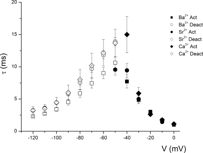
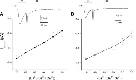
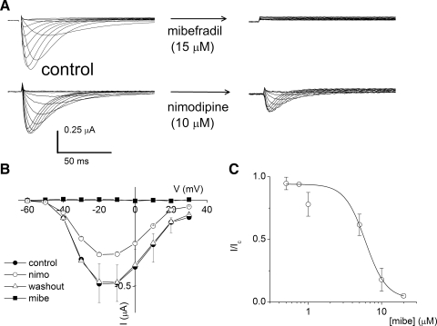
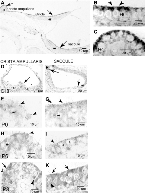
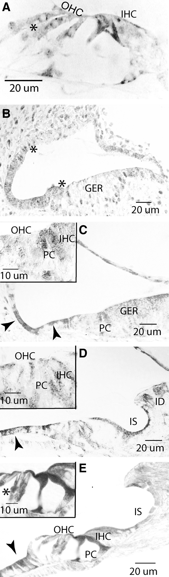
The molecular identity of non-Cav1.3 channels in auditory and vestibular hair cells has remained obscure, yet the evidence in support of their roles to promote diverse Ca2+-dependent functions is indisputable. Recently, a transient Cav3.1 current that serves as a functional signature for the development and regeneration of hair cells has been identified in the chicken basilar papilla. The Cav3.1 current promotes spontaneous activity of the developing hair cell, which may be essential for synapse formation. Here, we have isolated and sequenced the full-length complementary DNA of a distinct isoform of Cav3.1 in the mouse inner ear. The channel is derived from alternative splicing of exon14, exon25A, exon34, and exon35. Functional expression of the channel in Xenopus oocytes yielded Ca2+ currents, which have a permeation phenotype consistent with T-type channels. However, unlike most multiion channels, the T-type channel does not exhibit the anomalous mole fraction effect, possibly reflecting comparable permeation properties of divalent cations. The Cav3.1 channel was expressed in sensory and nonsensory epithelia of the inner ear. Moreover, there are profound changes in the expression levels during development. The differential expression of the channel during development and the pharmacology of the inner ear Cav3.1 channel may have contributed to the difficulties associated with identification of the non-Cav1.3 currents.


Figures

FIG. 1.
FIG. 1.
Reverse transcription–polymerase chain reaction (RT-PCR) detection of T-type Ca2+ subunit Cav3.1 channels in the mouse inner ear. A: RT-PCR products of Cav3.1 from cochlea complementary (c)DNA and utricle cDNA were separated on a 2% agarose gel with DNA Cav1.3 as positive control. M, DNA molecular weight marker; 1.3, Cav1.3 channel; 3.1, Cav3.1 channel; NTC, no template control. BD: alternative splicing of Cav3.1 channel in mice. B: 3 different transcripts derived from alternative combinations of the inclusion (+) and exclusion (−) of exon8, exon14, exon25a, exon34, and exon35 in Cav3.1. Gray boxes: constitutive exons; white boxes: alternative exons. Sizes of exons and introns are not drawn to scale. C: the schematic presentation of 3 Cav3.1 channel proteins showing the differences resulting from alternative splicing. The white cylinder, dashed line, and white line show the regions encoded by exon8, exon14, and exon25b, which are alternatively utilized, included or excluded in the mouse Cav3.1 channel isoforms. D: amino acid sequences of the regions encoded by alternative exons (underlined) in Cav3.1 channels.
FIG. 2.
FIG. 2.
Inner ear Cav3.1 channel currents. A: examples of inner ear Cav3.1 currents expressed in Xenopus oocytes. The current traces were recorded by applying hyperpolarizing and depolarizing voltage steps (step potentials were from −120 to 10 mV from a holding potential of −110 mV). Ba2+ was used as the charge carrier and the concentrations of the bath solution are indicated. B: the current–voltage (IV) relations of the peak current were plotted as a function of voltage. Changes in the magnitude of the current were observed as the external Ba2+ concentration was increased. A shift in the profile of the IV relation toward positive potentials as the external Ba2+ concentration was increased is consistent with increased surface charge screening effects of the divalent cation. C: the current magnitudes of the Cav3.1 channel were determined using different concentrations of Ba2+, Ca2+, and Sr2+ (2–65 mM). The solid lines represent fits with a Langmuir isotherm of the form I = Imax/(1 + KD/[ion]), where I and Imax represent the whole cell current and maximum current, respectively, and [ion] is the divalent ion concentration. The estimated apparent KD (in mM) values for the Cav3.1 were: 4.6 ± 0.6, 4.2 ± 0.4, and 3.1 ± 0.3 (n = 6), for Ba2+, Ca2+, and Sr2+, respectively.
FIG. 3.
FIG. 3.
Steady-state activation and inactivation of Cav3.1 currents. A: comparison of current records from oocytes injected with messenger (m)RNA of Cav3.1 channel in the presence of 10 mM Ba2+ (left) and Sr2+ (right) as permeant ions. Membrane currents were recorded at various command potentials from a holding potential of −110 mV in 10 mM divalent cation solutions. B: mean peak IV relation of Ba2+, Ca2+, and Sr2+ currents (n = 14). The magnitude of the currents invariably followed the order ISr > ICa > IBa. C: steady-state activation curves were plotted using the ratio of the conductance (g/gmax) against the step potentials. The conductance (g) was estimated from the equation g = I/(VErev), where Erev is the calculated reversal potential of the current. The continuous curves were generated from the Boltzmann function {g/gmax = [1 + exp(V1/2V)/Km]}−1. V1/2 is the half-activation voltage and Km is the slope factor. The estimated values of V1/2 and Km for Ba2+, Ca2+, and Sr2+ currents were (in mV): −33.2 ± 0.6 and 6.4 ± 0.5; −35.5 ± 0.6 and 5.6 ± 0.5; and −38.5 ± 0.6 and 6.3 ± 0.6 (n = 17), respectively. D: voltage-dependent inactivation of Ba2+, Ca2+, and Sr2+ currents. Twin-pulse protocol was elicited and the relative current at the test pulse (−10 mV) was plotted against the prepulse potentials. A 2-ms gap was allowed between the prepulse and test pulse for recovery from deactivation. The solid curves were generated from the Boltzmann function {I/Imax = [1 + exp(VV1/2)/Kh]}−1, where V1/2 is the half-inactivation voltage and Kh is the slope factor. The estimated values of V1/2 and Kh for Ba2+, Ca2+, and Sr2+ currents were (in mV): −52.1 ± 0.6 and 5.4 ± 0.5; −53.8 ± 0.6 and 5.5 ± 0.5; and −56.8 ± 0.5 and 4.7 ± 0.4 (n = 14), respectively. Error bars represent mean ± SD. The dotted curves represent the steady-state activation. Prominent window conductances can be seen from approximately −60 to −40 mV, with the peaks of the Ba2+, Ca2+, and Sr2+ occurring at (in mV) −42, −50, and −45, respectively.
FIG. 4.
FIG. 4.
Time constant of activation and deactivation of Cav3.1 currents. Scatterplots of the time constants of activation (solid symbols) and deactivation (open symbols) of Ba2+ (▪, □), Ca2+ (♦, ⋄), and Sr2+ (, ○) currents at various potentials.
FIG. 5.
FIG. 5.
Kinetics of inactivation of Cav3.1 currents. A: to estimate the time dependence of recovery from inactivation, we measured the recovery kinetics at different potentials using standard recovery time protocol as depicted in the inset. Examples of traces of Ba2+ currents used to determine the rate of recovery are also shown. We measured the amplitude of Ba2+ currents by depolarizing to a fixed potential, after variable amounts of time at the test potentials (as indicated). The amplitude of these currents was normalized to the amplitude of the currents activated at the initial test voltage, then plotted against the duration of steps to the test voltage. The kinetics of recovery from inactivation required at least 2 time constants (τ values) to accurately reflect the time course of Ba2+ current inactivation. The exponential fits to the data were: τ values (in ms) at −60 mV: 8.6 ± 1.6, 550.0 ± 24.4 (n = 9); at −80 mV: 79.5 ± 9.5, 292.6 ± 53.3 ms (n = 12); at −100 mV: 78.4 ± 5.5, 511.3 ± 61.5 (n = 9); at −120 mV: 58.6 ± 4.8, 395.8 ± 35.7 (n = 9). B: we examined the time dependence of development of inactivation after varying durations at a test potential. The time course of development of inactivation at the test potential and the τ values of development of inactivation were determined for the current traces (see inset for an example of the protocol and the corresponding Ba2+ current traces). One τ was sufficient to fit the time course of development of inactivation. The fits to the data were: τ values (in ms) −40 mV; 110.5 ± 9.3 (n = 7), −60 mV; 726.6 ± 51.9 (n = 7).
FIG. 6.
FIG. 6.
Permeation properties of Cav3.1 in the inner ear. Anomalous mole fraction effect (AMFE) is absent in Cav3.1. Peak tail currents were measured in mixtures of Ca2+ and Ba2+, in which the concentration of divalent ions was kept constant at 2.5 mM. RNA-injected oocytes were held at −110 mV, stepped to −30 mV, and the tail currents were measured at −40 mV (see insets A and B). A: no AMFE was observed in 2.5 mM Ca2+/Ba2+ (n = 7). Cav3.1 currents increased monotonically as a function of the Ba2+ mole fraction. The absence of an AMFE was observed at all tested voltages (−70 to −20 mV). B: using 2.5 mM Ba2+/Sr2+, and applying similar protocols, evaluation of the tail currents vs. the fraction produces a monotonic increase in tail currents as a function of the Ba2+ mole fraction (n = 7).
FIG. 7.
FIG. 7.
Pharmacology of Cav3.1 current. A: current traces were recorded from Cav3.1 RNA-injected oocytes. They were held at a holding potential of −110 mV and stepped to different depolarizing voltages. Ba2+ was the charge carrier in these experiments. For the example shown, after application of 15 μM mibefradil (mibe), the current was completely blocked. Additionally, 10 μM nimodipine reduced the current magnitude by about 2-fold. B: the IV relationship of currents in magnitude, showing the effects of 15 μM mibefradil and 10 μM nimodipine on control currents and current recovery after washout. C: mibefradil block of the transient Ba2+ current was dose dependent. The half-blocking concentration of mibefradil was estimated to be 5.8 ± 0.6 μM (n = 7).
FIG. 8.
FIG. 8.
Single-channel currents of Cav3.1 expressed in Xenopus oocytes. A: representative and consecutive single-channel traces recorded in a cell-attached patch with a pipette ([Ba+], 20 mM). The bath solution contained 140 mM K+ and the resting potential of oocytes was about 0 mV. The holding potential was −120 mV and the step potentials are indicated. B: an example of an amplitude histogram used to generate IV relationships at −90 mV. The IV relationships are shown in C and D for Ba2+ and Ca2+ currents, respectively. The single-channel conductances were: Ba2+, 10.7 ± 0.9 pS (n = 5) and Ca2+, 13.3 ± 1.1 pS (n = 6). C, closed; O, open.
FIG. 9.
FIG. 9.
Cav3.1 expression in the vestibular system. A: in the adult, Cav3.1 is expressed in all vestibular organs (arrow) and, to a lesser degree, in the underlying connective tissue (asterisk). B and C: higher-power views of the saccule (B) and ampulla (C) show staining in the apical portion of support cells and hair cells (B, between arrowheads). D and E: Cav3.1 is first noted at embryonic day 18 (E18) in all epithelial (asterisk) and neuroepithelial cells (arrows) in the membranous labyrinth. F and G: at postnatal day 0 (P0), the cytosol of macular and support cells (asterisk) showed light reactivity. H and I: by P6, a moderate immunoreactivity can be seen in the cytosol of hair cells (arrowheads) and support cells. The nerve chalice at the type I hair cells is negative (asterisk). J and K: by P8, moderately dark reactivity was seen in apices of hair cells and support cells (arrows). The type I perinuclear region shows light reactivity (arrowhead), whereas the afferent nerve chalice is negative.
FIG. 10.
FIG. 10.
Cav3.1 expression in the cochlear system. A: in the adult cochlea, punctuate staining is noted in the perinuclear region of the outer hair cells (OHCs). The Deiter's cup (asterisk) under each OHC stained weakly for Cav3.1, as did the cytosol of the inner hair cell (IHC). B: developmentally, expression of Cav3.1 is first noted at E18 in the membranous labyrinth region that will evolve into the OHCs supporting cells of the organ of Corti and the outer sulcus cells of the spiral ligament (between asterisks). C: at P0, expression of Cav3.1 has increased to a moderate intensity in the developing outer sulcus cells and supporting cells of the organ of Corti (arrowheads). A light punctate stain is seen in the pillar cells (PCs) and the greater epithelial ridge (GER). The inset shows light stain in the IHC region, but lack of reactivity in the OHCs. D: by P6, the inner sulcus (IS) epithelial cells and interdental (ID), cells in addition to the supporting cells (arrowhead), showed moderate reactivity. A light punctuate stain was detected at the plasmalemma of the IHCs and OHCs (see inset). E: at P8, a light-moderate reactivity was noted in the inner sulcus (IS) and supporting cells (arrowhead), whereas a more intense Cav3.1 expression exists in the pillar cells. The inset shows the reactivity in the cytosol of the hair cells, pillar cells, and the cup region of the Deiter cells (asterisk).
FIG. 11.
FIG. 11.
Cav3.1 in nonsensory tissues. A: the basal turn of the cochlea revealed moderate immunopositivity for Cav3.1 in fibrocytes and root cells of the spiral ligament (Sp Lig) (asterisks). Unlike the ID cells, the upper fibrocytes of the spiral limbus (white asterisk) showed strong reactivity with less intense staining noted in the lower spiral limbal fibrocytes (arrows). B: higher magnification of the spiral ligament revealed moderate positivity in lower type I fibrocytes (arrow) with less reactivity in the upper type I fibrocytes underlying the stria vascularis (StV). C: basal cells (BCs) of the stria vascularis (arrows) showed moderate reactivity. D: as positive control, we show strong reactivity for Cav3.1 in the Purkinje (P) layer of the cerebellum (arrows; Nahm et al. 2005). E: substitution of nonimmune serum and/or antigenic peptide for the Cav3.1 antibody failed to show any reactivity in the crista ampullaris (asterisk). OHC, outer hair cell; IHC, inner hair cell; IC, intermediate cell; MC, marginal cell; DC, dark cell.

Similar articles

Cited by

References

    1. Art JJ, Fettiplace R. Variation of membrane properties in hair cells isolated from the turtle cochlea. J Physiol 385: 207–242, 1987. - PMC - PubMed
    1. Balkowiec A, Katz DM. Cellular mechanisms regulating activity-dependent release of native brain-derived neurotrophic factor from hippocampal neurons. J Neurosci 22: 10399–10407, 2002. - PMC - PubMed
    1. Bao H, Wong WH, Goldberg JM, Eatock RA. Voltage-gated calcium channel currents in type I and type II hair cells isolated from the rat crista. J Neurophysiol 90: 155–164, 2003. - PubMed
    1. Barish ME, Mansdorf NB. Development of intracellular calcium responses to depolarization and to kainate and N-methyl-d-aspartate in cultured mouse hippocampal neurons. Brain Res Dev Brain Res 63: 53–61, 1991. - PubMed
    1. Bertolesi GE, Jollimore CA, Shi C, Elbaum L, Denovan-Wright EM, Barnes S, Kelly ME. Regulation of alpha1G T-type calcium channel gene (CACNA1G) expression during neuronal differentiation. Eur J Neurosci 17: 1802–1810, 2003. - PubMed

Publication types

MeSH terms

LinkOut - more resources