. 2008 Aug 27;28(35):8756-64.
doi: 10.1523/JNEUROSCI.2645-08.2008.

Developmental excitation of corticothalamic neurons by nicotinic acetylcholine receptors

Affiliations

Developmental excitation of corticothalamic neurons by nicotinic acetylcholine receptors

Sameera M Kassam et al. J Neurosci. .

Abstract

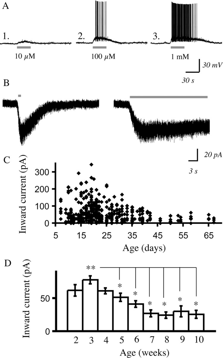
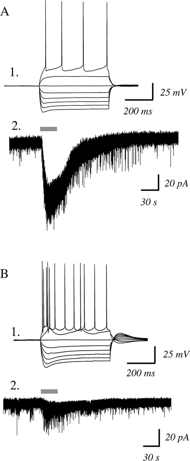
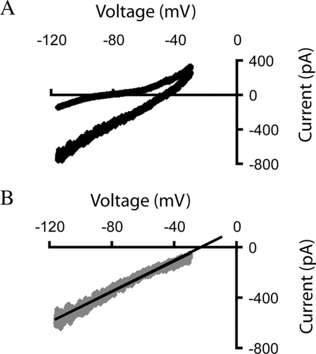
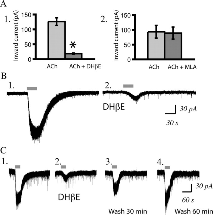
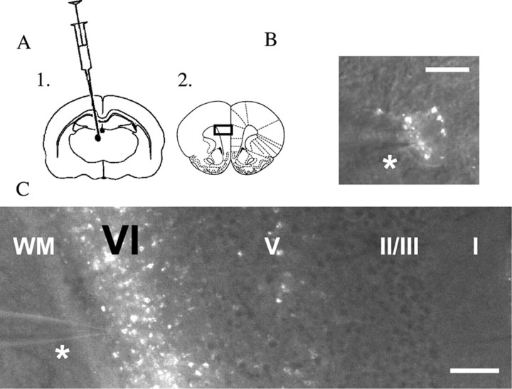
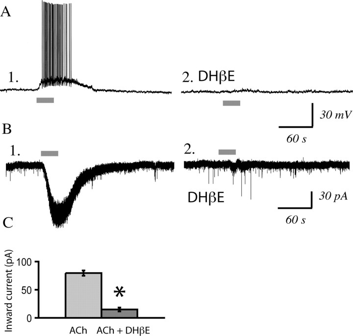
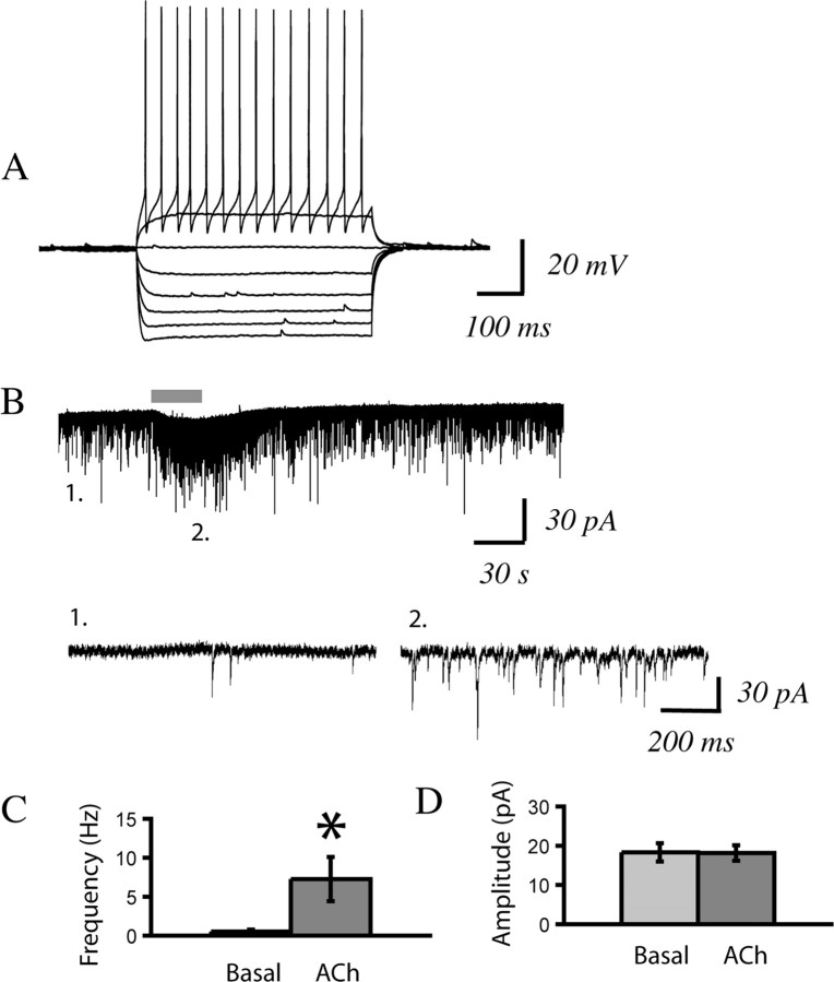
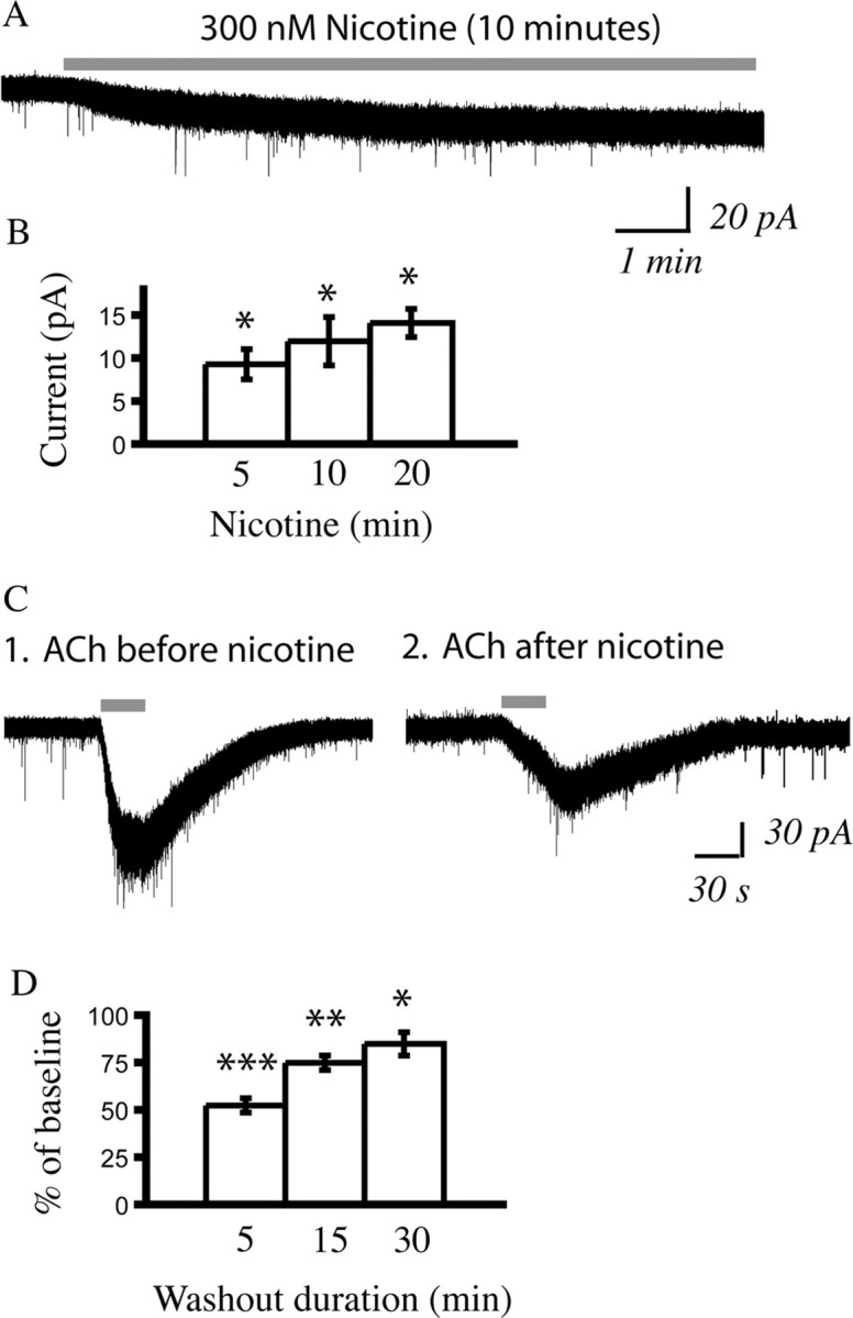
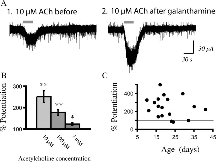
In this study, we show robust nicotinic excitation of pyramidal neurons in layer VI of prefrontal cortex. This layer contains the corticothalamic neurons, which gate thalamic activity and play a critical role in attention. Our experiments tested nicotinic excitation across postnatal development, using whole-cell recordings in prefrontal brain slices from rats. These experiments showed that layer VI neurons have peak nicotinic currents during the first postnatal month, a time period of intensive cortical development in rodents. We demonstrate that these currents are mediated directly by postsynaptic nicotinic receptors and can be suppressed by a competitive antagonist of alpha(4)beta(2)* nicotinic receptors. To record from identified corticothalamic neurons, we performed stereotaxic surgery to label the neurons projecting to medial dorsal thalamus. As hypothesized, recordings from these retrogradely labeled neurons in layer VI showed prominent nicotinic currents. Finally, we examined the effects of the drug nicotine on layer VI neurons and probed for the potential involvement of the accessory subunit, alpha(5), in their receptors. A level of nicotine similar to that found in the blood of smokers elicits a stable inward current in layer VI neurons, yet this exposure desensitizes approximately 50% of the subsequent current elicited by acetylcholine. An allosteric modulator of alpha(4)beta(2)alpha(5) receptors resulted in a 2.5-fold potentiation of submaximal nicotinic currents. This result is consistent with the expression of the relatively rare alpha(5) nicotinic subunit in layer VI. In summary, we show that layer VI corticothalamic neurons can be strongly excited during development by an unusual subtype of nicotinic receptor.


Figures

Figure 1.
Figure 1.
Nicotinic receptor stimulation with acetylcholine excites pyramidal neurons in layer VI in the presence of atropine to block muscarinic receptors. A2–A3, The concentration–response relationship for nicotinic depolarization of a layer VI pyramidal neuron from a P19 rat in current clamp (acetylcholine: 30 s, 5 min washout between sweeps; atropine: 200 nm present in all experiments). B, Voltage-clamp traces show that similar peak inward currents result from rapid local application of acetylcholine (1 mm, 1 s) and bath application (1 mm, 30 s). C, The scattergram shows peak inward currents from early postnatal development to adulthood elicited by acetylcholine (1 mm, 30 s). D, Summary data from the scattergram in C showing mean inward current elicited by acetylcholine in layer VI pyramidal neurons by postnatal week. The Kruskal–Wallis ANOVA shows a highly significant developmental effect (**p < 0.0001) in which the mean inward current at postnatal week 3 is significantly higher than the mean inward current in postnatal weeks 5 through 10 (*p < 0.05), as shown by Mann–Whitney tests.
Figure 2.
Figure 2.
Regular-spiking, rather than bursting, layer VI neurons are more likely to have large nicotinic inward currents. A1, Current-clamp recording showing membrane potential changes in response to injecting depolarizing and hyperpolarizing current steps in a regular firing neuron from a P33 rat. A2, A voltage-clamp trace from this neuron showing a robust inward current with acetylcholine (1 mm, 30 s) in the presence of atropine (200 nm). B1, This current-clamp recording is of a burst firing neuron from the same P33 rat. B2, A voltage-clamp trace from this neuron showing minimal inward current with acetylcholine (1 mm, 30 s) in the presence of atropine (200 nm).
Figure 3.
Figure 3.
Nicotinic currents elicited by acetylcholine are mediated by somatodendritic nicotinic receptors on layer VI neurons. A, The current–voltage curves in TTX (2 μm) for a layer VI neuron at baseline (upper curve) and in the presence of acetylcholine (1 mm, 30 s; lower curve, which appears thicker because of increased current noise). B, The current–voltage relationship is shown, and the reversal potential was found for the nicotinic current in this neuron from a P21 rat. This reversal potential is consistent with the equilibrium potential for a nonselective cationic channel under our recording conditions.
Figure 4.
Figure 4.
Layer VI pyramidal neurons are excited by α4β2* nicotinic acetylcholine receptors. A1, Bar chart showing that the competitive α4β2* antagonist DHβE (3 μm, 10 min) suppressed the nicotinic inward current [acetylcholine (ACh) (n = 20; *p < 0.00001, paired t test; age range examined: P7–P65). A2, Bar chart showing that the α7 receptor antagonist MLA (10 nm, 10 min) does not significantly suppress this current (n = 11; p = NS, paired t test; age range examined: P7–P50). B1, B2, Voltage-clamp traces showing the effect of acetylcholine (1 mm, 30 s) before and after DHβE (3 μm, 10 min) in a neuron from a P19 rat. C1–C4, Voltage-clamp traces in a neuron from a P21 rat showing the baseline effect of acetylcholine (C1), suppression with DHβE (3 μm, 10 min) (C2), partial recovery after 30 min washout (C3), and full recovery after 60 min washout of DHβE (C4) (note that the inward current is the same as in C1, but there is a flurry of sEPSCs near the peak that cannot be resolved on this scale).
Figure 5.
Figure 5.
Corticothalamic neurons are labeled by stereotaxic surgery to infuse the retrograde tracer rhodamine microspheres in medial dorsal thalamus. A1, A2, Schematics show the infusion location in the thalamus (A1) as well as the coronal prefrontal slice (A2), where layer VI medial prefrontal neurons are recorded 3–12 d postoperatively. The black rectangle on the coronal slice shows the location of the low-magnification image in C. B, A high-magnification image of a labeled corticothalamic neuron during recording (the patch pipette is just above the asterisk). Scale bar, 20 μm. C, A low-magnification image of this same neuron and pipette (above asterisk) showing the laminar location of the recorded neuron within medial prefrontal cortex. Note the strong retrograde labeling in layer VI [the layer adjacent to white matter (WM)] as well as the sparse labeling of the small population of corticothalamic neurons in layer V. Scale bar, 240 μm.
Figure 6.
Figure 6.
Identified corticothalamic neurons show robust nicotinic currents that are sensitive to the α4β2* antagonist, DHβE. A1, A2, Current-clamp trace showing the effects of acetylcholine (1 mm, 30 s) before and after DHβE (3 μm, 10 min) in a labeled corticothalamic neuron from a P21 rat. B1, B2, Voltage-clamp trace of another labeled neuron showing the effects of acetylcholine (1 mm, 30 s) before and after DHβE (3 μm, 10 min) in a P29 rat. C, Bar chart summarizing nicotinic acetylcholine (ACh) currents in labeled layer VI corticothalamic neurons before and after DHβE (n = 12; paired t test, *p < 0.000001).
Figure 7.
Figure 7.
As hypothesized, fast-spiking interneurons in layer VI show a prominent indirect effect of nicotinic acetylcholine stimulation. A, Current-clamp trace showing membrane potential changes in response to depolarizing and hyperpolarizing current steps, with action potentials that are characteristic of a fast-spiking interneuron. B1, B2, Voltage-clamp trace showing the effects of a 30 s application of acetylcholine (1 mm). The inset magnifies the EPSCs under baseline conditions (B1) and in the presence of acetylcholine (B2). C, Bar chart summarizing the mean change in spontaneous EPSC frequency with nicotinic stimulation (n = 14; *p < 0.05, paired t test). D, Bar chart summarizing the mean change in spontaneous EPSC amplitude with nicotinic stimulation (n = 14; p = NS, paired t test).
Figure 8.
Figure 8.
A persistent inward current is elicited by nicotine, which washes out rapidly but leaves a lingering suppression of the maximal effects of acetylcholine. A, A voltage-clamp trace showing a small but persistent inward current elicited in a neuron from a P19 rat by application of the drug nicotine (300 nm, 10 min). This concentration of nicotine is consistent with the peak blood level of nicotine seen in smokers (Henningfield et al., 1993). B, The bar chart showing the mean currents elicited by three different durations of nicotine application: 5 min (n = 31), 10 min (n = 25), and 20 min (n = 7) applications. All of the inward currents are significantly different when compared with baseline: *p < 0.001, paired t tests. The age range examined is P9-P41. C1, A voltage-clamp trace from a P21 rat showing a robust depolarization with acetylcholine (1 mm, 30 s) preceding the application of nicotine. C2, A voltage-clamp trace from the same neuron taken 5 min after the end of a 40 min nicotine application shows that the depolarization elicited by acetylcholine is decreased. D, Bar chart showing the highly significant suppression of the inward current elicited by acetylcholine (1 mm, 30 s; n = 31; ***p < 0.00001, paired t test) examined 5 min after nicotine application (300 nm, 5–10 min). This bar chart also shows the continued suppression of the responses of the neurons to acetylcholine at 15 min of washout (n = 11; **p < 0.01, paired t test) and 30 min of washout (n = 10; *p < 0.05, paired t test).
Figure 9.
Figure 9.
The nicotinic currents in layer VI can be dramatically potentiated by galanthamine, suggesting the involvement of α5-containing α4β2 nicotinic receptors. A, Voltage-clamp traces showing that the inward current elicited by acetylcholine (ACh) (10 μm, 30 s) is more than doubled after application of galanthamine (1 μm, 10 min) in a recording from a P10 rat. B, The bar chart shows how much different concentrations of acetylcholine are potentiated above baseline after application of galanthamine (0.1–1 μm). The left bar shows the potentiation for 10 μm acetylcholine (n = 18; **p < 0.001, paired t test), the middle bar shows potentiation for 100 μm acetylcholine (n = 23; **p < 0.001, paired t test), and the right bar shows potentiation for 1 mm acetylcholine (n = 19; *p < 0.01, paired t test). C, The scattergram shows the percent that a submaximal concentration of acetylcholine is potentiated by galanthamine (0.1–1 μm) over the age range examined: P10–P41. The gray line indicates the level at which the initial current was not altered by galanthamine.

References

    1. Belluardo N, Mudò G, Blum M, Cheng Q, Caniglia G, Dell'Albani P, Fuxe K. The nicotinic acetylcholine receptor agonist (+/−)-epibatidine increases FGF-2 mRNA and protein levels in the rat brain. Brain Res Mol Brain Res. 1999;74:98–110. - PubMed
    1. Berrettini W, Yuan X, Tozzi F, Song K, Francks C, Chilcoat H, Waterworth D, Muglia P, Mooser V. Alpha-5/alpha-3 nicotinic receptor subunit alleles increase risk for heavy smoking. Mol Psychiatry. 2008;13:368–373. - PMC - PubMed
    1. Bierut LJ, Stitzel JA, Wang JC, Hinrichs AL, Grucza RA, Xuei X, Saccone NL, Saccone SF, Bertelsen S, Fox L, Horton WJ, Breslau N, Budde J, Cloninger CR, Dick DM, Foroud T, Hatsukami D, Hesselbrock V, Johnson EO, Kramer J, Kuperman S, Madden PA, Mayo K, Nurnberger J, Jr, Pomerleau O, Porjesz B, Reyes O, Schuckit M, Swan G, Tischfield JA, Edenberg HJ, Rice JP, Goate AM. Variants in nicotinic receptors and risk for nicotine dependence. Am J Psychiatry. 2008 in press. - PMC - PubMed
    1. Breese CR, Lee MJ, Adams CE, Sullivan B, Logel J, Gillen KM, Marks MJ, Collins AC, Leonard S. Abnormal regulation of high affinity nicotinic receptors in subjects with schizophrenia. Neuropsychopharmacology. 2000;23:351–364. - PubMed
    1. Brody AL, Mandelkern MA, London ED, Olmstead RE, Farahi J, Scheibal D, Jou J, Allen V, Tiongson E, Chefer SI, Koren AO, Mukhin AG. Cigarette smoking saturates brain alpha 4 beta 2 nicotinic acetylcholine receptors. Arch Gen Psychiatry. 2006;63:907–915. - PMC - PubMed

Publication types

MeSH terms