

Mononuclear myeloid-derived "suppressor" cells express RAE-1 and activate natural killer cells
- PMID: 18753637
- PMCID: PMC2582006
- DOI: 10.1182/blood-2008-03-143776
Mononuclear myeloid-derived "suppressor" cells express RAE-1 and activate natural killer cells
Abstract
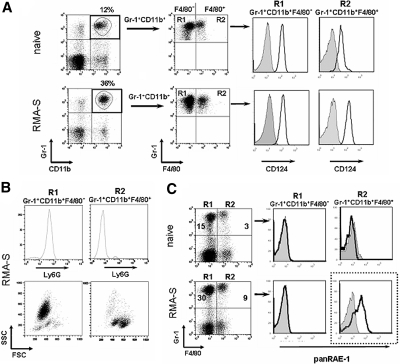
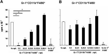
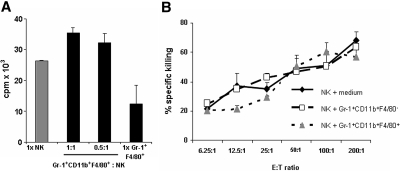
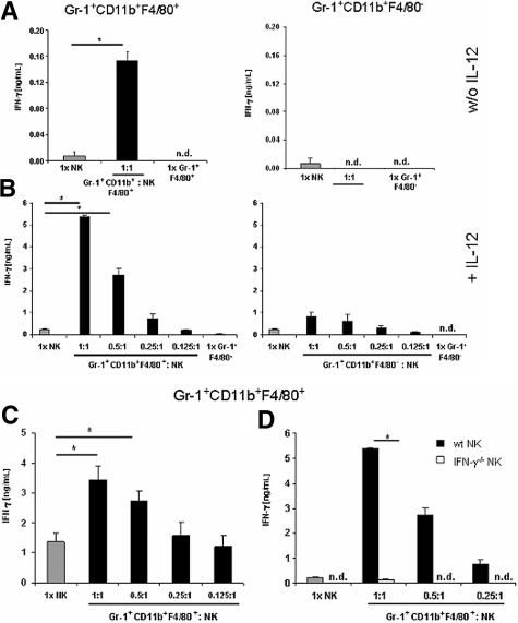
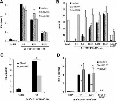
Myeloid-derived suppressor cells (MDSCs) accumulate in cancer patients and tumor-bearing mice and potently suppress T-cell activation. In this study, we investigated whether MDSCs regu-late natural killer (NK)-cell function. We discovered that mononuclear Gr-1(+)CD11b(+)F4/80(+) MDSCs isolated from RMA-S tumor-bearing mice do not suppress, but activate NK cells to produce high amounts of IFN-gamma. Gr-1(+)CD11b(+)F4/80(+) MDSCs isolated from tumor-bearing mice, but not myeloid cells from naive mice, expressed the ligand for the activating receptor NKG2D, RAE-1. NK-cell activation by MDSCs depended partially on the interaction of NKG2D on NK cells with RAE-1 on MDSCs. NK cells eliminated Gr-1(+)CD11b(+)F4/80(+) MDSCs in vitro and upon adoptive transfer in vivo. Finally, depletion of Gr-1(+) cells that comprise MDSCs confirmed their protective role against the NK-sensitive RMA-S lymphoma in vivo. Our study reveals that MDSCs do not suppress all aspects of antitumor immune responses and defines a novel, unexpected activating role of MDSCs on NK cells. Thus, our results have great impact on the design of immune therapies against cancer aiming at the manipulation of MDSCs.
Figures
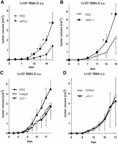
 ), isotype-matched control mAb (□), a neutralizing anti–IFN-γ mAb (A, ■), a depleting anti-NK1.1 mAb (B, ■), or an anti–Gr-1 mAb (C and D, ▴) and were inoculated subcutaneously with either RMA-S (A,B,C) or RMA lymphoma cells (D). (A-D) Tumor growth was monitored and is presented as mean plus or minus SD. Asterisks indicate statistical significance of P < .01, determined by a one-way analysis of variance (ANOVA) test. One representative experiment of at least 2 independent experiments is shown.
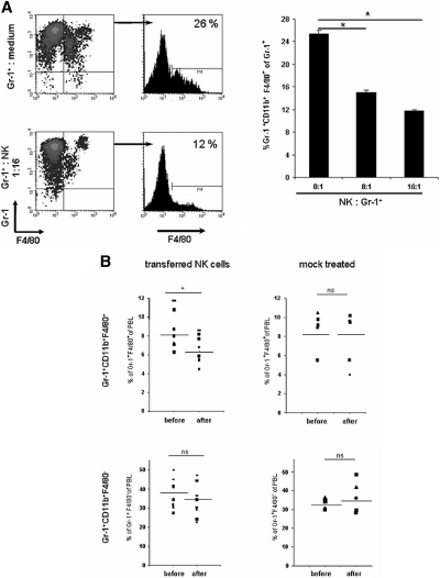
Similar articles
-
Activated NK cells reprogram MDSCs via NKG2D-NKG2DL and IFN-γ to modulate antitumor T-cell response after cryo-thermal therapy.J Immunother Cancer. 2022 Dec;10(12):e005769. doi: 10.1136/jitc-2022-005769. J Immunother Cancer. 2022. PMID: 36521929 Free PMC article.
-
Inhibition of tumor-derived prostaglandin-e2 blocks the induction of myeloid-derived suppressor cells and recovers natural killer cell activity.Clin Cancer Res. 2014 Aug 1;20(15):4096-106. doi: 10.1158/1078-0432.CCR-14-0635. Epub 2014 Jun 6. Clin Cancer Res. 2014. PMID: 24907113
-
Myeloid-derived Suppressor Cells Activate Liver Natural Killer Cells in a Murine Model in Uveal Melanoma.Curr Med Sci. 2022 Oct;42(5):1071-1078. doi: 10.1007/s11596-022-2623-3. Epub 2022 Oct 17. Curr Med Sci. 2022. PMID: 36245024
-
Myeloid Derived Suppressor Cells Interactions With Natural Killer Cells and Pro-angiogenic Activities: Roles in Tumor Progression.Front Immunol. 2019 Apr 18;10:771. doi: 10.3389/fimmu.2019.00771. eCollection 2019. Front Immunol. 2019. PMID: 31057536 Free PMC article. Review.
-
The NKG2D/NKG2DL Axis in the Crosstalk Between Lymphoid and Myeloid Cells in Health and Disease.Front Immunol. 2018 Apr 23;9:827. doi: 10.3389/fimmu.2018.00827. eCollection 2018. Front Immunol. 2018. PMID: 29740438 Free PMC article. Review.
Cited by
-
Mechanisms of immune suppression by myeloid-derived suppressor cells: the role of interleukin-10 as a key immunoregulatory cytokine.Open Biol. 2020 Sep;10(9):200111. doi: 10.1098/rsob.200111. Epub 2020 Sep 16. Open Biol. 2020. PMID: 32931721 Free PMC article. Review.
-
Evaluation of innate and adaptive immunity contributing to the antitumor effects of PD1 blockade in an orthotopic murine model of pancreatic cancer.Oncoimmunology. 2016 Mar 16;5(6):e1160184. doi: 10.1080/2162402X.2016.1160184. eCollection 2016 Jun. Oncoimmunology. 2016. PMID: 27471630 Free PMC article.
-
Mouse tumor vasculature expresses NKG2D ligands and can be targeted by chimeric NKG2D-modified T cells.J Immunol. 2013 Mar 1;190(5):2455-63. doi: 10.4049/jimmunol.1201314. Epub 2013 Jan 25. J Immunol. 2013. PMID: 23355740 Free PMC article.
-
Myeloid derived suppressor cells in human diseases.Int Immunopharmacol. 2011 Jul;11(7):802-7. doi: 10.1016/j.intimp.2011.01.003. Epub 2011 Jan 13. Int Immunopharmacol. 2011. PMID: 21237299 Free PMC article. Review.
-
Tumor-derived CSF-1 induces the NKG2D ligand RAE-1δ on tumor-infiltrating macrophages.Elife. 2018 May 14;7:e32919. doi: 10.7554/eLife.32919. Elife. 2018. PMID: 29757143 Free PMC article.
References
-
- Serafini P, Borrello I, Bronte V. Myeloid suppressor cells in cancer: recruitment, phenotype, properties, and mechanisms of immune suppression. Semin Cancer Biol. 2006;16:53–65. - PubMed
-
- Huang B, Pan PY, Li Q, et al. Gr-1+CD115+ immature myeloid suppressor cells mediate the development of tumor-induced T regulatory cells and T-cell anergy in tumor-bearing host. Cancer Res. 2006;66:1123–1131. - PubMed
Publication types
MeSH terms
Substances
LinkOut - more resources
Full Text Sources
Other Literature Sources
Molecular Biology Databases
Research Materials
