. 2009 Jun;83(6):609-17.
doi: 10.1007/s00204-008-0352-4. Epub 2008 Aug 27.

Protective effects of curcumin against gamma radiation-induced ileal mucosal damage

Affiliations

Protective effects of curcumin against gamma radiation-induced ileal mucosal damage

Meryem Akpolat et al. Arch Toxicol. 2009 Jun.

Abstract

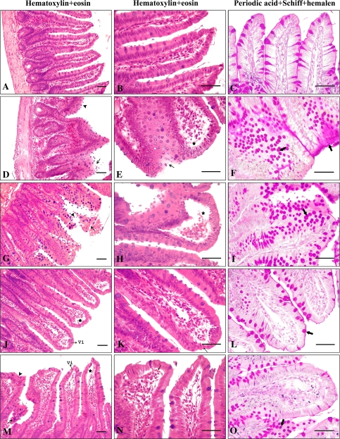
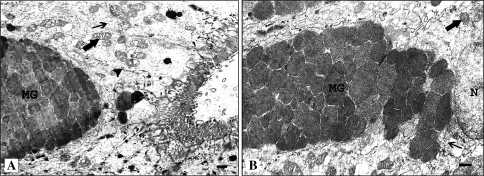
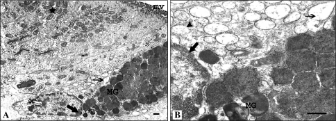
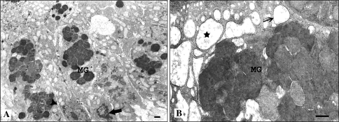
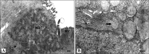
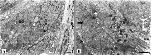
The major objective of this study was to test curcumin as a potential radioprotectant for the ileum goblet cells of the rat. Wistar albino rats were used in the study. Group A was the control group and group B was the single dose radiation group. Group C was the two dose radiation group (4 days interval). The rats in groups D and E were given a daily dose of 100 mg/kg of curcumin for 14 and 18 days, respectively. During the curcumin administration period, the rats in group D were exposed to abdominal area gamma (gamma)-ray dose of 5 Gy on the 10th day and group E was exposed to same dose radiation on the 10th and 14th day. Irradiation and treatment groups were decapitated on the 4th day after exposure to single or two-dose irradiation and ileum tissues were removed for light and electron microscopic investigation. Single or two dose 5 Gy gamma-irradiation caused a marked intestinal mucosal injury in rats on the 4th day. Radiation produced increases in the number of goblet cells. Curcumin appears to have protective effects against radiation-induced damage, suggesting that clinical transfer is feasible.


Figures

Fig. 1
Fig. 1
Histological examination of rat small intestine taken from the segments of ileum. Photomicrographs of ileum sections stained with H + E (a, b, d, e, g, h, j, k, m, n), and PAS + Hl (c, f, i, l, o). a–c Control rats ileum; showing normal morphology. d–f Single dose radiation-treated rats; shortened and thickened villi (arrowhead) and degenerative changes in the epithelial cells (arrow), lifting of epithelial layer from the lamma propria (asterisks), and increase in the goblet cells and mucins showing strongly positive to PAS staining (thick arrows). g–i Two dose radiation-treated rats; shortened and irregular villi (arrowhead) and breaking in the epithelial cells (arrow), massive subepithelial lifting and capillary congestion in the villus (asterisks), and increase in the goblet cells and mucins showing strongly positive to PAS staining (thick arrows). j–l Single dose radiation-treated with curcumin rats; villi were generally normal (Vi) although apical regions of some villi were lightly subepithelial lifting (asterisks) and thinning (arrow), normal scattered goblet cells (thick arrows). m–o Two dose radiation-treated with curcumin rats; development of subepithelial space (asterisks) and lightly capillary congestion (thick arrows) usually at the apex of the villus, occasional occurrence of thickened villi (arrowhead) along with normal villi (Vi) and breaking in the epithelial cells (arrow), goblet cells showing strongly positive to PAS staining (thick arrows) (scale bar: 50 μm)
Fig. 2
Fig. 2
Electron micrographs of goblet cells in the Liberkühn cryptal and villus region of control rat ileum (a, b). The mucus granules (MG) have a typical honeycomb shape with well-packed in goblet cell. The cytoplasm of epithelial cells has cisternae of ER (arrow). Mitochondria (thick arrows) have a typical cylindrical shape with organized cristae. Cell contact region (arrowhead) and oval shape nucleus (N) (uranyl acetate and lead citrate, scale bar 0.5 μm)
Fig. 3
Fig. 3
Electron micrographs of single dose radiation-treated rat ileum (a, b). The dilatation of ER cisternae (arrow) and degranulation of RER membranes (arrowhead). Marginal condensation of chromatin onto the nuclear lamina (thick arrows) and invagination of the nuclear envelope (x). Condensed mucus granules (MG). Increase in mitochondria (asteriks) and shortening in the microvillous of enterocytes (mv) (uranyl acetate and lead citrate, scale bar 0.5 μm)
Fig. 4
Fig. 4
Electron micrographs of two dose radiation-treated rat ileum (a, b). The massive dilatation of ER cisternae (asteriks) and degranulation of RER membranes (arrow). Marginal condensation of chromatin onto the nuclear lamina (thick arrows). Abnormal spread and condensation of mucus granules (MG) in goblet cell. Separation cell contact region (arrowhead) (uranyl acetate and lead citrate, scale bar 0.5 μm)
Fig. 5
Fig. 5
Electron micrographs of single dose radiation-treated with curcumin rat ileum show little evidence of damaged cellular structure (a, b). The mucus granules (MG) have a typical honeycomb shape with well-packed in goblet cell. The goblet cell has regular cisternae of RER (arrow). Mitochondria (thick arrows) have a typical cylindrical shape with organized cristae. Microvilli have uniform shape (Mv) (uranyl acetate and lead citrate, scale bar 0.2 μm)
Fig. 6
Fig. 6
Electron micrographs of two dose radiation-treated with curcumin rat ileum (a, b). The goblet cell has regular cisternae of RER (arrow). Invagination of the nuclear envelope (x) and slightly spread of mucus granules (MG) in goblet cell. Mitochondria (thick arrows) have a typical cylindrical shape with organized cristae. Microvilli have a lightly irregular shape (Mv). Cell contact regions have a lightly disintegration (arrowhead). Lysosome (L) (uranyl acetate and lead citrate, scale bar 0.5 μm)

Similar articles

Cited by

References

    1. {'text': '', 'ref_index': 1, 'ids': [{'type': 'DOI', 'value': '10.2307/3579813', 'is_inner': False, 'url': 'https://doi.org/10.2307/3579813'}, {'type': 'PubMed', 'value': '9806614', 'is_inner': True, 'url': 'https://pubmed.ncbi.nlm.nih.gov/9806614/'}]}
    2. Barcellos-Hoff MH (1998) How the tissue respond to damage at the cellular level? The role of cytekines in irradiated tissues. Radiat Res 150:109–120 - PubMed
    1. {'text': '', 'ref_index': 1, 'ids': [{'type': 'DOI', 'value': '10.3109/02841868509134403', 'is_inner': False, 'url': 'https://doi.org/10.3109/02841868509134403'}, {'type': 'PubMed', 'value': '2994383', 'is_inner': True, 'url': 'https://pubmed.ncbi.nlm.nih.gov/2994383/'}]}
    2. Becciolini A, Fabbrica D, Cremonini D, Balzi M (1985) Quantitative changes in the goblet cells of the rat small intestine after irradiation. Acta Radiol Oncol 24:291–299 - PubMed
    1. {'text': '', 'ref_index': 1, 'ids': [{'type': 'DOI', 'value': '10.1111/j.1365-2184.1997.tb00932.x', 'is_inner': False, 'url': 'https://doi.org/10.1111/j.1365-2184.1997.tb00932.x'}, {'type': 'PMC', 'value': 'PMC6496338', 'is_inner': False, 'url': 'https://pmc.ncbi.nlm.nih.gov/articles/PMC6496338/'}, {'type': 'PubMed', 'value': '9375028', 'is_inner': True, 'url': 'https://pubmed.ncbi.nlm.nih.gov/9375028/'}]}
    2. Becciolini A, Balzi M, Fabbrica D, Potten CS (1997) The effects of irradiation at different times of the day on rat intestinal goblet cells. Cell Prolif 30(3–4):161–170 - PMC - PubMed
    1. {'text': '', 'ref_index': 1, 'ids': [{'type': 'DOI', 'value': '10.1080/095530098141942', 'is_inner': False, 'url': 'https://doi.org/10.1080/095530098141942'}, {'type': 'PubMed', 'value': '9690687', 'is_inner': True, 'url': 'https://pubmed.ncbi.nlm.nih.gov/9690687/'}]}
    2. Brennan PC, Carr KE, Seed T, McCullough JS (1998) Acute and protracted radiation effects on small intestinal morphological parameters. Int J Radiat Biol 73:691–698 - PubMed
    1. {'text': '', 'ref_index': 1, 'ids': [{'type': 'PubMed', 'value': '6750767', 'is_inner': True, 'url': 'https://pubmed.ncbi.nlm.nih.gov/6750767/'}]}
    2. Carr KE (1981) Scanning electron microscopy of tissue response to irradiation. Scann Electron Microsc 4:35–46 - PubMed

Publication types