. 2008 Sep 2;52(10):843-50.
doi: 10.1016/j.jacc.2008.05.043.

Dysregulation of antioxidant mechanisms contributes to increased oxidative stress in calcific aortic valvular stenosis in humans

Affiliations

Dysregulation of antioxidant mechanisms contributes to increased oxidative stress in calcific aortic valvular stenosis in humans

Jordan D Miller et al. J Am Coll Cardiol. .

Abstract

Objectives: The aim of this study was to determine whether oxidative stress is increased in calcified, stenotic aortic valves and to examine mechanisms that might contribute to increased oxidative stress.

Background: Oxidative stress is increased in atherosclerotic lesions and might play an important role in plaque progression and calcification. The role of oxidative stress in valve disease is not clear.

Methods: Superoxide (dihydroethidium fluorescence and lucigenin-enhanced chemiluminescence), hydrogen peroxide H2O2 (dichlorofluorescein fluorescence), and expression and activity of pro- and anti-oxidant enzymes were measured in normal valves from hearts not suitable for transplantation and stenotic aortic valves that were removed during surgical replacement of the valve.

Results: In normal valves, superoxide levels were relatively low and distributed homogeneously throughout the valve. In stenotic valves, superoxide levels were increased 2-fold near the calcified regions of the valve (p < 0.05); noncalcified regions did not differ significantly from normal valves. Hydrogen peroxide levels were also markedly elevated in calcified regions of stenotic valves. Nicotinamide adenine dinucleotide phosphate oxidase activity was not increased in calcified regions of stenotic valves. Superoxide levels in stenotic valves were significantly reduced by inhibition of nitric oxide synthases (NOS), which suggests uncoupling of the enzyme. Antioxidant mechanisms were reduced in calcified regions of the aortic valve, because total superoxide dismutase (SOD) activity and expression of all 3 SOD isoforms was significantly decreased. Catalase expression also was reduced in pericalcific regions.

Conclusions: This study provides the first evidence that oxidative stress is increased in calcified regions of stenotic aortic valves from humans. Increased oxidative stress is due at least in part to reduction in expression and activity of antioxidant enzymes and perhaps to uncoupled NOS activity. Thus, mechanisms of oxidative stress differ greatly between stenotic aortic valves and atherosclerotic arteries.


Figures

Figure 1
Figure 1. Superoxide Levels Detected With DHE Fluorescence and Lucigenin-Enhanced Chemiluminescence
Superoxide (red fluorescence) in a normal (A) and stenotic (B) aortic valve detected with dihydroethidine (DHE) fluorescence. Superoxide levels were markedly elevated near the calcified (calc) region of the valve and were markedly reduced by the addition of polyethylene glycol superoxide dismutase. Magnified images of noncalcified (non-calc) and calcified regions of a stenotic valve with DHE staining are shown in C and D. (E) Superoxide levels measured with lucigenin-enhanced chemiluminescence in corresponding regions of normal and stenotic valves (n = 14 control valves, n = 20 stenotic valves; *p < 0.05 vs. noncalcified stenotic tissue; #p < 0.05 vs. base and tip of normal valves).
Figure 2
Figure 2. H2O2 Detected With DCF Fluorescence
Hydrogen peroxide (H2O2) in a normal (A) and stenotic (C) aortic valve detected with dichlorofluorescein (DCF) fluorescence. Levels of H2O2 were markedly elevated near the calcified regions of the valve, and most of the DCF fluorescence was eliminated by pre-incubation of the slide with polyethylene glycol (PEG)-catalase (CAT) (B and D). (E) The PEG-CAT–inhibitable fraction of DCF fluorescence in normal (base and tip regions) and stenotic (calcified and noncalcified regions) aortic valves (n = 4 normal valves, n = 7 stenotic valves; *p < 0.05 vs. noncalcified stenotic tissue, #p < 0.05 vs. base region of normal valves). Abbreviations as in Figure 1.
Figure 3
Figure 3. SOD Expression and Activity in Normal and Stenotic Aortic Valves
(A) Expression of the 3 superoxide dismutase (SOD) isoforms in normal and noncalcified and calcified regions of stenotic aortic valves (n = 16 normal valves, n = 15 stenotic valves). (B) Regional total SOD activity in stenotic aortic valves (n = 10 normal valves, n = 11 stenotic valves; *p < 0.05 vs. normal valves; #p < 0.05 vs. noncalcified stenotic tissue). mRNA = messenger ribonucleic acid; other abbreviations as in Figure 1.
Figure 4
Figure 4. Catalase Expression in Normal Valves and Noncalcified and Calcified Regions of Stenotic Aortic Valves
n = 10 normal valves, n = 11 stenotic valves; *p < 0.05 versus normal valves; #p < 0.05 versus noncalcified stenotic tissue. Abbreviations as in Figures 1 and 3.
Figure 5
Figure 5. Pro-Oxidative Enzymes in Normal and Stenotic Aortic Valves
(A) Expression of subunits of nicotinamide adenine dinucleotide phosphate (NADPH) oxidase in normal (n = 16) and stenotic aortic valves (n = 15 valves, with 15 noncalcified and 15 calcifed regions). (B) The NADPH oxidase activity in normal (n = 8 base and tip regions) and stenotic aortic valves (n = 14 calcified and noncalcified regions). Effects of inhibitors of enzymatic sources of superoxide in noncalcified (C) and calcified (D) regions of stenotic aortic valves measured with lucigenin-enhanced chemiluminescence (n = 5 to 11/group). APO = apocynin; DPI = diphenylidonium; RLU = relative light units; other abbreviations as in Figures 1 and 3.
Figure 6
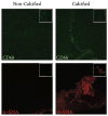
Figure 6. Macrophage and Activated Myofibroblast Markers in a Stenotic Human Valve
Antibodies against CD68 and α-SMA were used to detect macrophages and activated myofibroblasts, respectively, in noncalcified and calcified regions. Representative images from n = 5. Inset images are negative control images from adjacent sections.
Figure 7
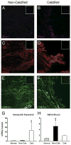
Figure 7. Immunofluorescent Staining in Noncalcified and Calcified Regions of Stenotic Aortic Valves
Immunofluorescent staining for Msx1 (A and B), Msx2 (C and D), and CBFA1 (E and F) in noncalcified and calcified regions of stenotic aortic valves (inset images are negative control images from adjacent sections; representative images from n = 5/stain). Panels G and H show messenger ribonucleic acid (mRNA) levels of osteopontin and CBFA1/Runx2 mRNA in normal valves and in noncalcified and calcified regions of stenotic aortic valves (*p < 0.05 vs. normal tissue, #p < 0.05 vs. noncalcified stenotic tissue, n = 8/group). Abbreviations as in Figure 1.
Figure 8
Figure 8. Different Mechanisms Generating Oxidative Stress in Vascular Lesions and Calcifed Stenotic Aortic Valves
↔ = no change; +/− = increases or decreases have been reported; NADPH = nicotinamide adenine dinucleotide phosphate; NOS = nitric oxide synthase.

Comment in

References

    1. Bonow RO, Carabello B, de Leon AC, et al. ACC/AHA guidelines for the management of patients with valvular heart disease: executive summary. A report of the American College of Cardiology/American Heart Association Task Force on Practice Guidelines (Committee on Management of Patients With Valvular Heart Disease) J Am Coll Cardiol. 1998;32:1486–588. - PubMed
    1. Lindroos M, Kupari M, Valvanne J, Strandberg T, Heikkila J, Tilvis R. Factors associated with calcific aortic valve degeneration in the elderly. Eur Heart J. 1994;15:865–70. - PubMed
    1. Messika-Zeitoun D, Bielak LF, Peyser PA, et al. Aortic valve calcification: determinants and progression in the population. Arterioscler Thromb Vasc Biol. 2007;27:642–8. - PubMed
    1. Otto CM, Kuusisto J, Reichenbach DD, Gown AM, O'Brien KD. Characterization of the early lesion of ‘degenerative’ valvular aortic stenosis. Histological and immunohistochemical studies. Circulation. 1994;90:844–53. - PubMed
    1. Rajamannan NM, Subramaniam M, Caira F, Stock SR, Spelsberg TC. Atorvastatin inhibits hypercholesterolemia-induced calcification in the aortic valves via the Lrp5 receptor pathway. Circulation. 2005;112:I229–34. - PMC - PubMed

Publication types