Randomized Controlled Trial
. 2008 Sep 2;52(10):869-81.
doi: 10.1016/j.jacc.2008.04.055. Epub 2008 Jul 2.

The peroxisome proliferator-activated receptor-gamma agonist pioglitazone represses inflammation in a peroxisome proliferator-activated receptor-alpha-dependent manner in vitro and in vivo in mice

Affiliations
Randomized Controlled Trial

The peroxisome proliferator-activated receptor-gamma agonist pioglitazone represses inflammation in a peroxisome proliferator-activated receptor-alpha-dependent manner in vitro and in vivo in mice

Gabriela Orasanu et al. J Am Coll Cardiol. .

Abstract

Objectives: Our aim was to investigate if the peroxisome proliferator-activated receptor (PPAR)-gamma agonist pioglitazone modulates inflammation through PPARalpha mechanisms.

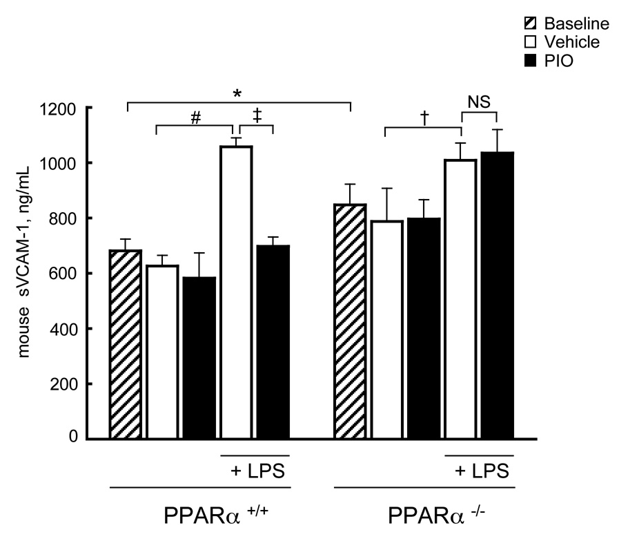
Background: The thiazolidinediones (TZDs) pioglitazone and rosiglitazone are insulin-sensitizing PPARgamma agonists used to treat type 2 diabetes (T2DM). Despite evidence for TZDs limiting inflammation and atherosclerosis, questions exist regarding differential responses to TZDs. In a double-blinded, placebo-controlled 16-week trial among recently diagnosed T2DM subjects (n = 34), pioglitazone-treated subjects manifested lower triglycerides and lacked the increase in soluble vascular cell adhesion molecules (sVCAM)-1 evident in the placebo group. Previously we reported PPARalpha but not PPARgamma agonists could repress VCAM-1 expression. Since both triglyceride-lowering and VCAM-1 repression characterize PPARalpha activation, we studied pioglitazone's effects via PPARalpha.

Methods: Pioglitazone effects on known PPARalpha responses--ligand binding domain activation and PPARalpha target gene expression--were tested in vitro and in vivo, including in wild-type and PPARalpha-deficient cells and mice, and compared with the effects of other PPARgamma (rosiglitazone) and PPARalpha (WY14643) agonists.

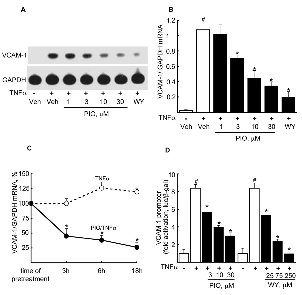
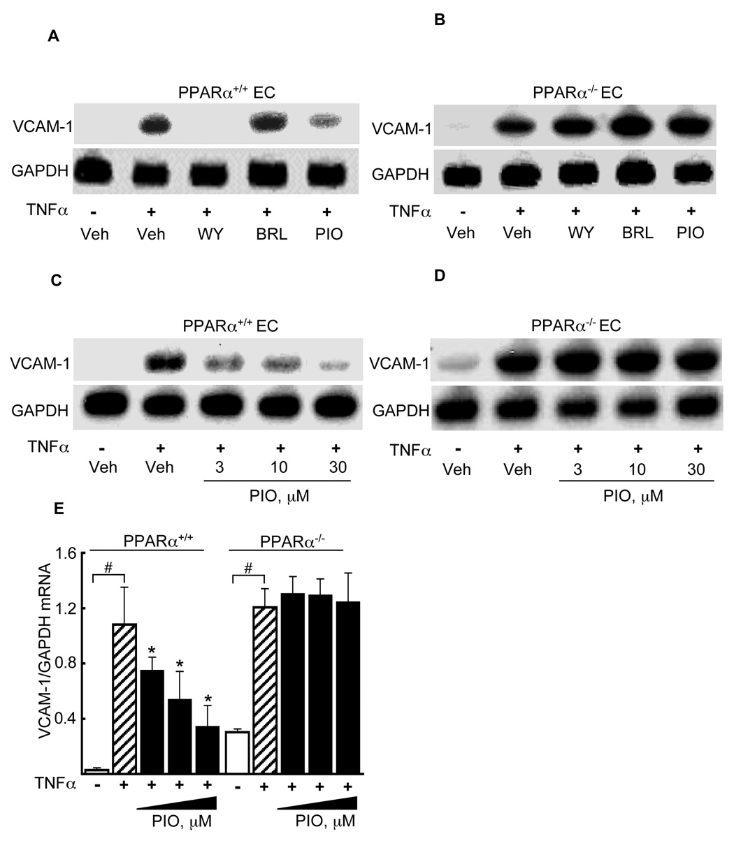
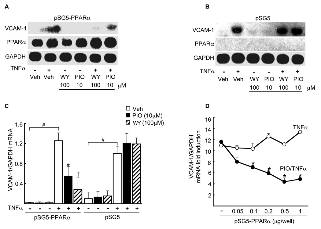
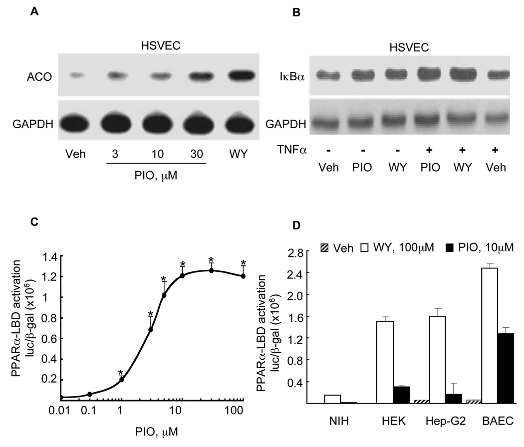
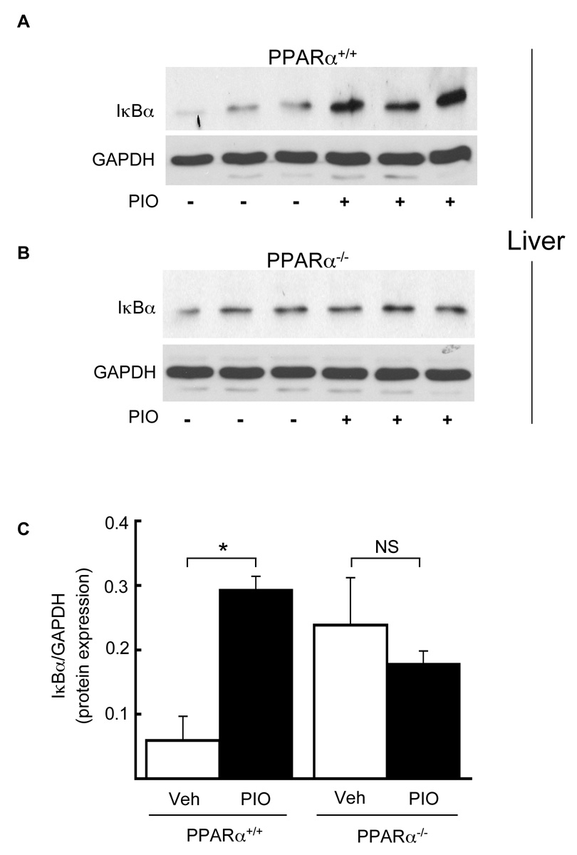
Results: Pioglitazone repressed endothelial TNFalpha-induced VCAM-1 messenger ribonucleic acid expression and promoter activity, and induced hepatic IkappaBalpha in a manner dependent on both pioglitazone exposure and PPARalpha expression. Pioglitazone also activated the PPARalpha ligand binding domain and induced PPARalpha target gene expression, with in vitro effects that were most pronounced in endothelial cells. In vivo, pioglitazone administration modulated sVCAM-1 levels and IkappaBalpha expression in wild-type but not PPARalpha-deficient mice.

Conclusions: Pioglitazone regulates inflammatory target genes in hepatic (IkappaBalpha) and endothelial (VCAM-1) settings in a PPARalpha-dependent manner. These data offer novel mechanisms that may underlie distinct TZD responses.


Figures

Figure 1
Figure 1. Pioglitazone reduces TNFα-induced VCAM-1 mRNA expression in a dose- and time-dependent manner in human saphenous vein ECs (HSVECs)
(A) Northern blot analysis of TNFα-induced VCAM-1 mRNA expression was performed on HSVECs pretreated in the absence or presence of pioglitazone (18 h) at the concentrations shown prior to TNFα stimulation (10 ng/mL, 10 h). The effects of the PPARα agonist WY14643 (100 µM) are provided for comparison. One representative Northern blot (n=3) is shown. (B). The effect of the pioglitazone concentrations on VCAM-1/GAPDH mRNA was quantified from the Northern blots above. (n=3, #p<0.05, TNFα-induced vs. vehicle, *p<0.05, pioglitazone/TNFα vs. TNFα alone, Mann-Whitney test). (C) The time dependent effects of pioglitazone exposure (10 µM) on TNFα-induced VCAM-1 expression was tested in HSVECs using Northern blotting. Results are shown as a percent of the TNFα effect alone at 3 h, mean ± SD (n = 3; *, p<0.05). (D) The effect of pioglitazone versus vehicle on the human VCAM-1 promoter transiently transfected into BAEC before TNFα stimulation are shown (left). For comparison, the effect of the PPARα agonist WY14643 on the VCAM-1 promoter is also shown (right). All responses were normalized to β-galactosidase (pCMV-β-Gal) (n = 3 per each treatment, #p<0.05 TNFα vs. vehicle; *p<0.05 pioglitazone or WY14643 vs. TNFα alone, Mann-Whitney test).
Figure 2
Figure 2. Pioglitazone represses TNFα-induced VCAM-1 expression in a PPARα-dependent manner
ECs isolated from PPARα+/+ (A) and PPARα−/− (B) mouse hearts were pretreated with WY14643, rosiglitazone (BRL), or pioglitazone at the concentrations shown (18 h) before mouse TNFα stimulation and subsequent Northern blotting for VCAM-1 mRNA and GAPDH expression. One representative blot of three is shown. Northern blotting for VCAM-1 expression was repeated in the presence of the dose range of pioglitazone shown in EC from PPARα+/+ (C) and PPARα−/− (D) mice. (E) Quantification of the effects of pioglitazone on VCAM-1 mRNA in PPARα+/+ and PPARα−/− EC relative to GAPDH mRNA expression (n = 3, #p<0.05 TNFα vs. vehicle; *p<0.05 pioglitazone/TNFα vs. TNFα alone, Mann-Whitney test).
Figure 3
Figure 3. PPARα is required for pioglitazone-mediated repression of TNFα induced endothelial VCAM-1 mRNA expression
Northern blot analysis was performed on total RNA isolated from PPARα−/− ECs transfected either with PPARα-containing pSG5 overexpression vector (A) or pSG5 alone (B) before stimulation with TNFα in either the absence or presence of WY (100 µM) or pioglitazone (10 µM), with subsequent probing for VCAM-1 or GAPDH expression. (C) Quantification of the VCAM-1 mRNA response to pioglitazone relative to GAPDH mRNA expression levels (n = 3, #p<0.05 TNFα vs. vehicle; *p<0.05 pioglitazone/TNFα vs. TNFα alone; p<0.05 WY14643/TNFα vs. TNFα alone, Mann-Whitney test). (D) Cells were transfected in (A, B) but with a concentration gradiant of pSG5-PPARα as shown before TNFα stimulation in either the absence or presence of pioglitazone 10 µM (n = 3, *p<0.05 pioglitazone/TNFα vs. TNFα alone).
Figure 4
Figure 4. Pioglitazone induces known PPARα target gene expression and PPARα–LBD activation in ECs
(A) Northern blot analysis in HSVECs was performed for the PPARα target gene acyl-CoA-oxidase (ACO) and compared to GAPDH in HSVEC pretreated (16 h) with pioglitazone or WY14643 at the concentrations shown before TNFα stimulation. B) Western blot analysis for IκBα expression was performed on total protein extracts (50µg) from HSVECs treated with either pioglitazone (10 µM) or WY14643 (250 µM) before stimulation with human TNFα. (C) Standard LBD activation assays were performed in BAECs stimulated with pioglitazone at the concentrations shown (0.01–100 µM). (D) PPARα-LBD assays were done as before but comparing responses in NIH/3T3 (fibroblasts), HEK293 (human kidney epithelial), Hep-G2 (hepatic) and BAEC cell lines before stimulation with pioglitazone or WY14643 (both 10 µM). Values are expressed as luciferase/β-Gal activity mean± SD (n = 3. *p<0.05 BAEC vs. NIH/3T3, ** vs. HEK293, *** vs. Hep-G2, both Student’s t and Mann-Whitney tests).
Figure 5
Figure 5. Pioglitazone induces IκBα protein expression in vivo in a PPARα dependent manner
PPARα+/+ (A) and PPARα−/− (B) mice were treated with pioglitazone (20mg/kg, 7 days via gavage) before livers were harvested and total protein extracted for Western blot analysis of IκBα and GAPDH protein levels. Each lane represents a single mouse. (C) The effects of pioglitazone (black bars) and vehicle (white bars) on IκBα protein expression were quantified and normalized to GAPDH expression. Mean values SD are shown (n = 4 mice/group. *p<0.05 pioglitazone vs. vehicle, Mann-Whitney test).
Figure 6
Figure 6. Pioglitazone decreases LPS-induced soluble VCAM-1 in PPARα+/+ but not PPARα−/− mice in vivo
PPARα+/+ and PPARα−/− mice were treated with pioglitazone or vehicle alone before LPS injection (n = 9/genotype as in Methods). sVCAM-1 levels in PPARα+/+ and PPARα−/− mice are shown at baseline (*p<0.007 PPARα−/− vs. PPARα+/+ mice) and after LPS injection in mice treated with either vehicle or pioglitazone (PPARα+/+, n=9, #p<0.002 LPS/vehicle vs. vehicle; p<0.01 pioglitazone/LPS vs. vehicle/LPS) and in PPARα−/− mice (n=9, p<0.05 vehicle/LPS vs. vehicle; (NS) non-significant pioglitazone/LPS vs. vehicle/LPS, significance determined using Mann-Whitney test). The mean serum sVCAM-1 concentration of each group ± SD is shown.

Comment in

References

    1. Glass CK, Witztum JL. Atherosclerosis. the road ahead. Cell. 2001:503–516. - PubMed
    1. Libby P. Current concepts of the pathogenesis of the acute coronary syndromes. Circulation. 2001:365–372. - PubMed
    1. Semenkovich CF. Insulin resistance and atherosclerosis. J Clin Invest. 2006:1813–1822. - PMC - PubMed
    1. Brown JD, Plutzky J. Peroxisome proliferator-activated receptors as transcriptional nodal points and therapeutic targets. Circulation. 2007:518–533. - PubMed
    1. Lindberg M, Astrup A. The role of glitazones in management of type 2 diabetes. A DREAM or a nightmare? Obes Rev. 2007:381–384. - PubMed

Publication types

MeSH terms