. 2008 Oct 15;586(20):4891-904.
doi: 10.1113/jphysiol.2008.156281. Epub 2008 Aug 28.

Epileptiform activities in slices of hippocampus from mice after intra-hippocampal injection of kainic acid

Affiliations

Epileptiform activities in slices of hippocampus from mice after intra-hippocampal injection of kainic acid

Caroline Le Duigou et al. J Physiol. .

Abstract

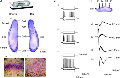
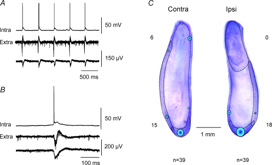
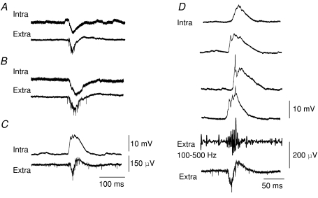
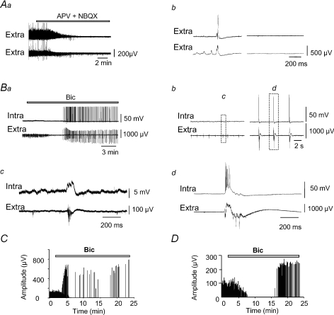
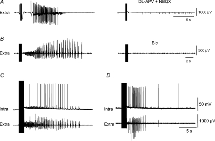
Intra-hippocampal kainate injection induces the emergence of recurrent seizures after a delay of 3-4 weeks. We examined the cellular and synaptic basis of this activity in vitro using extracellular and intracellular records from longitudinal hippocampal slices. These slices permitted recordings from the dentate gyrus, the CA3 and CA1 regions and the subiculum of both the injected and the contralateral non-injected hippocampus. A sclerotic zone was evident in dorsal regions of slices from the injected hippocampus, while ventral regions and tissue from the contralateral hippocampus were not sclerotic. Interictal field potentials of duration 50-200 ms were generated spontaneously in both ipsilateral and contralateral hippocampal slices, but not in the sclerotic region, at 3-12 months after injection. They were initiated in the CA1 and CA3 regions and the subiculum. They were blocked by antagonists at glutamatergic receptors and were transformed into prolonged epileptiform events by GABAergic receptor antagonists. The membrane potential and the reversal potential of GABAergic synaptic events were more depolarized in CA1 pyramidal cells from kainate-treated animals than in control animals. Ictal-like events of duration 8-80 s were induced by tetanic stimulation (50 Hz, 0.2-1 s) preferentially in dorsal contralateral and ventral ipsilateral slices. Similar events were initiated by focal application of a combination of high K(+) and GABA. These data show that both interictal and ictal-like activities can be induced in slices of both ipsilateral and contralateral hippocampus from kainate-treated animals and suggest that changes in cellular excitability and inhibitory synaptic signalling may contribute to their generation.


Figures

Figure 1
Figure 1. Characteristics of longitudinal hippocampal slices
A, Nissl-stained sections of contralateral and ipsilateral hippocampus at 7 months after kainate injection. Sclerosis of the CA1 and CA3 regions and a dispersion of dentate granule cells are evident in the dorsal part of the ipsilateral slice. The CA1 and CA3 regions as well as the dentate gyrus (GD) and subiculum (Sub) are indicated. Aa and Ab show the transition between the CA1 region and subiculum indicated by the squares in A with a double staining for Nissl substance (blue) and immunostaining for Calbindin (brown), selectively expressed by CA1 pyramidal cells. The inset above shows schematically how slices were cut. B, responses of two subicular cells (Ba, Bb) and a CA1 pyramidal cell (Bc) to depolarizing and hyperpolarizing current injections. Subicular cells fired either repetitively or with an initial burst. CA1 cells fired repetitively. C, simultaneous extracellular records showing the spread of synchronous firing along the subiculum in response to stimulation at a dorsal CA1 site in the presence of 20 μm bicuculline. The distance between extracellular recording electrodes was about 500 μm and the stimulus site was 150 μm distant from the first electrode.
Figure 2
Figure 2. Sites of generation and form of interictal activity
A and B, intracellular record from a CA1 pyramidal cell and correlated extracellular interictal-like activity at two sites in the CA1 region at different time scales. C, summary of initiation sites for interictal-like events. The diameter of circles represents the probability that interictal activity was generated at that site. In 6 out of 39 slices, activity was initiated in dorsal contralateral hippocampus, in 15 slices in ventral contralateral hippocampus and in the ventral part of ipsilateral slices by 18 of 39 slices.
Figure 3
Figure 3. Intracellular correlates of interictal-like activity
A, a cell that received a sequence of excitatory and inhibitory synaptic events; B, a cell that received purely hyperpolarizing events; and C, purely excitatory events during interictal field potentials. Membrane potentials were −72, −72 and −67 mV, respectively. D, variability of intracellular behaviour for a cell receiving exclusively depolarizing synaptic events. Note the fast hyperpolarizing deflections superimposed on the depolarizing envelope of the second, third and fourth intracellular traces. These events were uncorrelated with rapid oscillations in the extracellular field potential (band-pass filter 100–500 Hz).
Figure 4
Figure 4. Effects of blocking excitation and inhibition on interictal-like activity
Aa, dl-APV (100 μm) and NBQX (10 μm) suppress interictal-like activity in extracellular records from the CA1 region of ventral contralateral hippocampus. Ab, expanded traces before and after antagonist application. Ba, effect of bicuculline (Bic, 20 μm). Intracellular and extracellular records from the ventral CA1 region of a contralateral slice. Interictal activity was suppressed. After a delay of about 2 min a distinct, synchronous activity emerged. Bb–d, expanded traces show events induced by bicuculline were larger and prolonged. Bc before: and Bd after bicuculline. C and D, the amplitude of field potentials plotted against time after bicuculline application. In C, synchronous events were not interrupted but their amplitude increased and frequency decreased. In D interictal synchrony was suppressed before larger events emerged after a delay.
Figure 5
Figure 5. Cellular and synaptic properties in KA-injected and control animals
A, voltage dependance of evoked synaptic responses recorded in presence of APV/NBQX for a CA1 cell near the initiation site of interictal-like events in ventral contralateral hippocampus. The synaptic potential reversed at −62 mV and resting potential was −70 mV. Ba, voltage dependance of IPSPs from a CA1 pyramidal cell of a KA-treated animal and, Bb, from a cell of a control animal. Ca shows a firing threshold of −55 mV and a resting membrane potential of −62 mV for a CA1 cell recorded from a KA-treated animal and Cb shows a cell from control animal with resting membrane potential −80 mV and firing threshold −59 mV. D, resting membrane potentials and reversal potentials for GABAergic synaptic events in neurones of epileptic (n= 9) and control animals (n= 9). Data from individual cells are joined with mean and s.e.m. values on either side. E, values of resting membrane potential and spike threshold in neurones from KA-treated (n= 22) and control animals (n= 7).
Figure 6
Figure 6. Ictal-like activities induced by tetanic stimulation
A, different patterns of ictal-like event induced by tetanic stimulation. In the upper two traces, extracellular records from CA1 and the subiculum, show a high-frequency activity followed by a sequence of population bursts. In the lower traces, both from the subiculum, ictal-like activity consisted of a series of population bursts of duration about 15 s. B, ictal-like activity in slices from control animals. Extracellular records from CA1 (upper) and the subiculum (lower) showed that events were of shorter duration and spread less far. C, sites at which tetanic stimulation induced an ictal-like activity. The diameter of circles represents the probability that ictal-like activity was induced: dorsal contralateral regions, 12 of 28 slices; ventral contralateral sites, 3 of 28 slices; ventral injected hippocampus, 14 of 27 slices. Ictal-like activity was induced most effectively at borders between the CA1 region and the subiculum or CA3. D, responses induced by different intensities of stimulation (at 50 Hz, 0.5 s). An intensity of 20 V induced an increase in multi-unit activity, at 30 V clonic-like population bursts were induced with a latency of about 2 s, and at 50 V a high-frequency activity was induced with a shorter latency before population bursts emerged.
Figure 7
Figure 7. Pharmacological properties and intracellular correlates of two forms of ictal activity
A, an ictal-like event induced by tetanic stimulation, consisting of an acceleration of clonic bursts, was suppressed by antagonists at glutamatergic receptors (NBQX, 10 μm and d,l-APV 100 μm). B, an ictal-like event with an initial fast, low voltage activity, which was suppressed by bicuculline (20 μm). C, simultaneous intracellular and field recordings from contralateral dorsal subiculum of an ictal event with an initial fast, low voltage activity followed by clonic bursts. D, intracellular and field records of an ictal-like event in CA1 which consisted of an acceleration of clonic bursts.
Figure 8
Figure 8. Focal application of high K+ together with GABA initiates ictal-like activity
A, focal application of 50 nl of GABA (100 μm in external solution) induced an increase in the frequency of multi-unit activity. B, focal application of 50 nl of high-K+ (50 mm) induced an increase in multi-unit activity. D, in the same slice, application of 100 nl GABA (100 μm) and K+ (50 mm) induced a larger increase in multi-unit activity that culminated in an ictal-like event of duration 45 s. A, B and C show extracellular traces above and the frequency of detected action potentials below. D, extracellular record of an ictal-like activity induced in the CA3 region of the same contralateral dorsal hippocampal slice by focal stimulation in stratum radiatum.

Similar articles

Cited by

References

    1. Andersson PB, Perry VH, Gordon S. The CNS acute inflammatory response to excitotoxic neuronal cell death. Immunol Lett. 1991;30:177–181. - PubMed
    1. Avoli M, Barbarosie M, Lucke A, Nagao T, Lopantsev V, Kohling R. Synchronous GABA-mediated potentials and epileptiform discharges in the rat limbic system in vitro. J Neurosci. 1996;16:3912–3924. - PMC - PubMed
    1. Becker AJ, Chen J, Zien A, Sochivko D, Normann S, Schramm J, Elger CE, Wiestler OD, Blumcke I. Correlated stage- and subfield-associated hippocampal gene expression patterns in experimental and human temporal lobe epilepsy. Eur J Neurosci. 2003;18:2792–2802. - PubMed
    1. Ben-Ari Y. Limbic seizure and brain damage produced by kainic acid: mechanisms and relevance to human temporal lobe epilepsy. Neuroscience. 1985;14:375–403. - PubMed
    1. Ben-Ari Y, Cossart R. Kainate, a double agent that generates seizures: two decades of progress. Trends Neurosci. 2000;23:580–587. - PubMed

Publication types