. 2008 Oct 15;586(20):4843-57.
doi: 10.1113/jphysiol.2008.154096. Epub 2008 Aug 28.

Regulation of smooth muscle contractility by the epithelium in rat vas deferens: role of ATP-induced release of PGE2

Affiliations

Regulation of smooth muscle contractility by the epithelium in rat vas deferens: role of ATP-induced release of PGE2

Ye Chun Ruan et al. J Physiol. .

Abstract

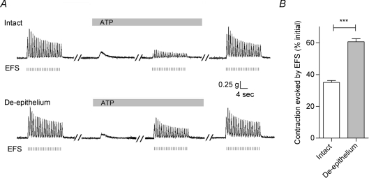
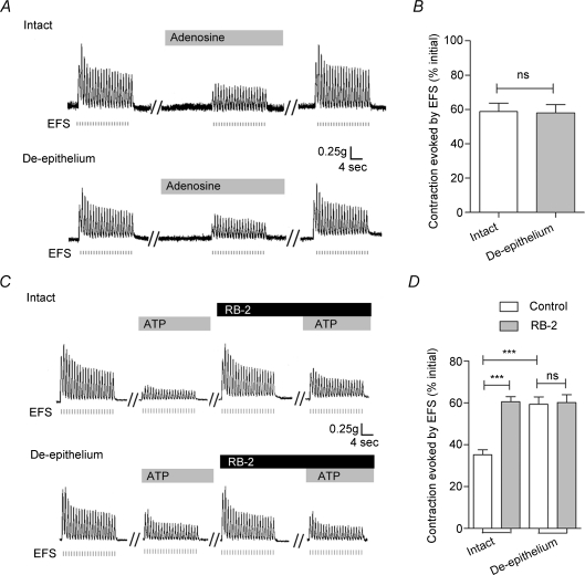
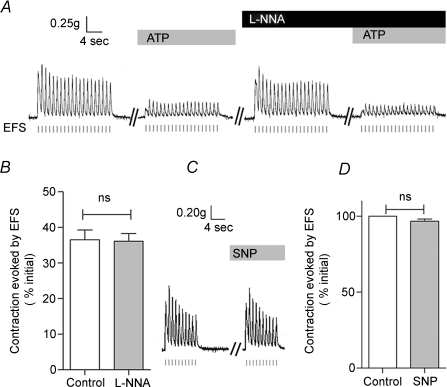
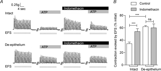
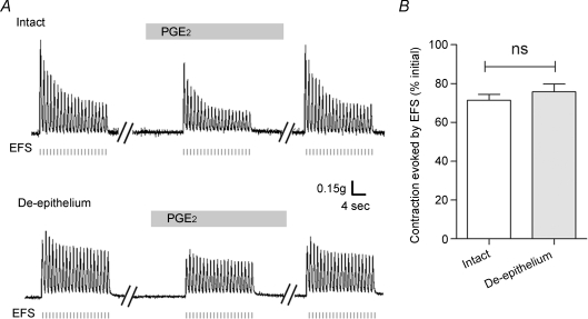
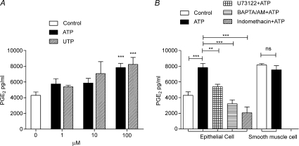
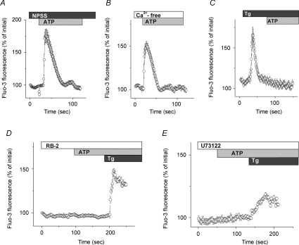
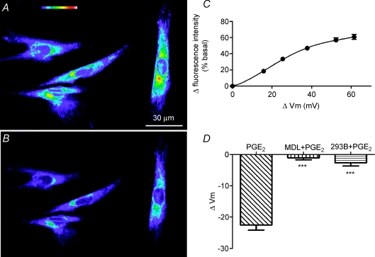
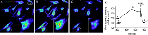
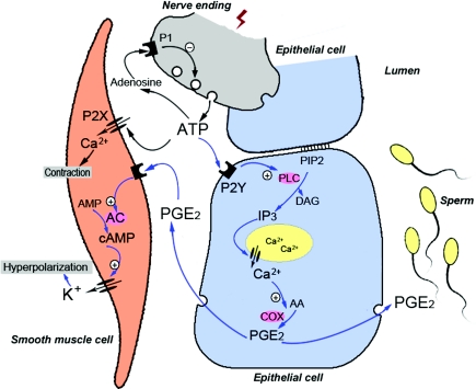
Recent studies suggest that the epithelium might modulate the contractility of smooth muscle. However, the mechanisms underlying this regulation are unknown. The present study investigated the regulation of smooth muscle contraction by the epithelium in rat vas deferens and the possible factor(s) involved. Exogenously applied ATP inhibited electrical field stimulation (EFS)-evoked smooth muscle contraction in an epithelium-dependent manner. As the effects of ATP on smooth muscle contractility were abrogated by inhibitors of prostaglandin synthesis, but not by those of nitric oxide synthesis, prostaglandins might mediate the effects of ATP. Consistent with this idea, PGE(2) inhibited EFS-evoked smooth muscle contraction independent of the epithelium, while ATP and UTP induced the release of PGE(2) from cultured rat vas deferens epithelial cells, but not smooth muscle cells. The ATP-induced PGE(2) release from vas deferens epithelial cells was abolished by U73122, an inhibitor of phospholipase C (PLC) and BAPTA AM, a Ca(2+) chelator. ATP also transiently increased [Ca(2+)](i) in vas deferens epithelial cells. This effect of ATP on [Ca(2+)](i) was independent of extracellular Ca(2+), but abolished by the P2 receptor antagonist RB2 and U73122. In membrane potential measurements using a voltage-sensitive dye, PGE(2), but not ATP, hyperpolarized vas deferens smooth muscle cells and this effect of PGE(2) was blocked by MDL12330A, an adenylate cyclase inhibitor, and the chromanol 293B, a blocker of cAMP-dependent K(+) channels. Taken together, our results suggest that ATP inhibition of vas deferens smooth muscle contraction is epithelium dependent. The data also suggest that ATP activates P2Y receptor-coupled Ca(2+) mobilization leading to the release of PGE(2) from epithelial cells, which in turn activates cAMP-dependent K(+) channels in smooth muscle cells leading to the hyperpolarization of membrane voltage and the inhibition of vas deferens contraction. Thus, the present findings suggest a novel regulatory mechanism by which the epithelium regulates the contractility of smooth muscle.


Figures

Figure 1
Figure 1. Epithelium dependence of the ATP-induced inhibition of the EFS-evoked contraction in rat vas deferens
A, representative mechanograms showing the inhibitory effect of exogenously applied ATP (100 μm) on EFS-evoked contractions of rat vas deferens preparations from the same animal with (upper trace) and without (lower trace) the epithelium. EFS pulses are indicated below the traces as short vertical bars. Bars above each mechanogram indicate when tissues are exposed to ATP. Breaks (5–30 min) in the duration of the mechanogram are indicated by oblique parallel lines. B, summary of results showing the effects of removal of the epithelium on the ATP (100 μm)-induced inhibition of EFS-evoked contractions. Data are expressed as the percentage of the initial response prior to ATP addition. Columns and error bars are mean +s.e.m. (n= 26; ***P < 0.001).
Figure 2
Figure 2. Effects of adenosine and RB2 on EFS-evoked contractions of rat vas deferens smooth muscle in the absence and presence of the epithelium
A, representative mechanograms show the inhibitory effects of exogenously applied adenosine (100 μm) on EFS-evoked contraction of rat vas deferens preparations with (upper trace) and without (lower trace) the epithelium. B, quantification of the effects of adenosine (100 μm) on EFS-evoked contractions. Data are means +s.e.m. (n= 6). C, representative mechanograms showing the EFS-evoked contraction of rat vas deferens preparations with (upper trace) and without (lower trace) the epithelium, before and after treatment with ATP (100 μm) and RB2 (100 μm). D, summary of results showing the effects of RB2 (100 μm) on the ATP-induced attenuation of EFS-evoked contractions in preparations with or without the epithelium. Data are mean +s.e.m. (n= 8; ***P < 0.001; ns: P > 0.05). Other details as in Fig. 1.
Figure 3
Figure 3. Nitric oxide (NO) does not mediate the ATP-induced inhibition of EFS-evoked contractions in rat vas deferens
A and B, representative mechanograms and summary data show the effects of l-NNA (300 μm) on the inhibition of EFS-evoked contractions by ATP (100 μm). Data are means +s.e.m. (n= 9; ns: P > 0.05, n= 9). C, D, representative mechanograms and summary data show the effects of SNP (100 μm) on EFS-evoked contractions. Data are means +s.e.m. (n= 5; ns: P > 0.05). Other details as in Fig. 1.
Figure 4
Figure 4. Indomethacin attenuates the ATP-induced inhibition of contraction in rat vas deferens
A, representative mechanograms of EFS-evoked contractions from rat vas deferens preparations with (upper trace) and without (lower trace) the epithelium, before and after treatment with ATP (100 μm) and indomethacin (10 μm). B, summary of results showing the effects of indomethacin (10 μm) on the ATP-induced inhibition of contraction in preparations with and without the epithelium. Data are mean +s.e.m. (n= 9; ***P < 0.001; ns: P > 0.05). Other details as in Fig. 1.
Figure 5
Figure 5. Effects of PGE2 on EFS-evoked contractions in rat vas deferens
A, representative mechanograms from preparations with (upper trace) and without (lower trace) the epithelium show the effects of PGE2 (1 μm) on the EFS-evoked contractions. B, summary of effects of PGE2 (1 μm) on EFS-evoked contractions in preparations with or without the epithelium. Data are mean +s.e.m. (n= 6; ns: P > 0.05). Other details as in Fig. 1.
Figure 6
Figure 6. Characterization of primary cultures of rat vas deferens smooth muscle and epithelial cells
A and B, bright field images of primary cultures of epithelial and smooth muscle cells of rat vas deferens. C and E, fluorescence images of epithelial cells showing FITC immunoreactivity for PCK and α-SMA, respectively. D and F, fluorescence images of smooth muscle cells showing FITC immunoreactivity for α-SMA and PCK, respectively.
Figure 7
Figure 7. ATP-induced PGE2 release from rat vas deferens epithelial cells
A, concentration-dependent effects of ATP and UTP on the release of PGE2 from cultured vas deferens epithelial cells. B, effects of BAPTA AM (100 μm), U73122 (10 μm) and indomethacin (10 μm) on the release of PGE2 from epithelial cells stimulated by ATP (100 μm). The effects of ATP (100 μm) on PGE2 release from smooth muscle cells is also shown. Data are means +s.e.m. (n= 3–4; **P < 0.01, ***P < 0.001, ns: P > 0.05).
Figure 8
Figure 8. ATP-induced increase in intracellular Ca2+ concentration in rat vas deferens epithelial cells
A and B, intracellular calcium transients elicited by ATP (100 μm) detected by fluo-3 fluorescence in normal physiological saline solution (A) and Ca2+-free physiological saline solution (B). CE, inhibitory effects of Tg (1 μm), RB2 (100 μm) and U73122 (10 μm) on the calcium transients elicited by ATP (100 μm). Data are expressed as a percentage of the initial fluo-3 fluorescence. Circles and error bars are mean ±s.e.m. (n= 17–25).
Figure 9
Figure 9. PGE2-induced hyperpolarization of rat vas deferens smooth muscle cells
A and B, fluorescence images of vas deferens smooth muscle cells loaded with the voltage-sensitive dye DiBAC(4)3 in the absence and presence of PGE2 (10 μm), respectively. Top of A, colour coding bar with increasing fluorescence intensity representing membrane potentials from strongly hyperpolarized (purple) to strongly depolarized (red to white). C, calibration curve with measured changes in fluorescence intensity (% of basal) as a function of membrane potential changes (calculated from changes in potassium gluconate concentrations). Data are mean ±s.e.m. (n= 15). Error bars are smaller than symbol size except where shown. D, summary of membrane potential changes (ΔVm) elicited by PGE2 alone and PGE2 and either MDL12330A (MDL, 10 μm) or the chromanol 293B (293B, 20 μm). Data are means +s.e.m. (PGE2, n= 24; MDL + PGE2, n= 22; 293B + PGE2, n= 19; ***P < 0.0001).
Figure 10
Figure 10. ATP-induced depolarization and PGE2-induced hyperpolarization of rat vas deferens smooth muscle cells
A, B and C are fluorescence images captured at times a, b and c in the time course D, respectively. The insets show higher resolution images of the circled cells. D, time course of fluorescence intensity in vas deferens smooth muscle cells showing the effects of ATP (100 μm) and PGE2 (10 μm). Arrow heads indicate the addition of ATP and PGE2. Similar results were observed in 12 other experiments.
Figure 11
Figure 11. Mechanisms underlying the multiple regulatory actions of ATP in the vas deferens
The schematic model shows communication between a neurone (grey), epithelial cells (blue) and a smooth muscle cell (pink). Blue arrows represent the signalling pathways or cellular events identified in the present study, whereas black arrows denote signalling pathways and cellular events described in previous studies of the vas deferens (Sneddon & Westfall, 1984; Driessen et al. 1994). See text for further information.

Comment in

Similar articles

Cited by

References

    1. Aitken RJ, Kelly RW. Analysis of the direct effects of prostaglandins on human sperm function. J Reprod Fertil. 1985;73:139–146. - PubMed
    1. Balzary RW, Cocks TM. Lipopolysaccharide induces epithelium- and prostaglandin E2-dependent relaxation of mouse isolated trachea through activation of cyclooxygenase (COX)-1 and COX-2. J Pharmacol Exp Ther. 2006;317:806–812. - PubMed
    1. Bos CL, Richel DJ, Ritsema T, Peppelenbosch MP, Versteeg HH. Prostanoids and prostanoid receptors in signal transduction. Int J Biochem Cell Biol. 2004;36:1187–1205. - PubMed
    1. Brauner T, Hulser DF, Strasser RJ. Comparative measurements of membrane potentials with microelectrodes and voltage-sensitive dyes. Biochim Biophys Acta. 1984;771:208–216. - PubMed
    1. Bucheimer RE, Linden J. Purinergic regulation of epithelial transport. J Physiol. 2004;555:311–321. - PMC - PubMed

Publication types

MeSH terms