. 2008 Oct;49(10):1407-16.
doi: 10.1093/pcp/pcn124. Epub 2008 Aug 28.

Separated transcriptomes of male gametophyte and tapetum in rice: validity of a laser microdissection (LM) microarray

Affiliations

Separated transcriptomes of male gametophyte and tapetum in rice: validity of a laser microdissection (LM) microarray

Keita Suwabe et al. Plant Cell Physiol. 2008 Oct.

Abstract

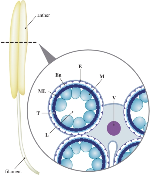
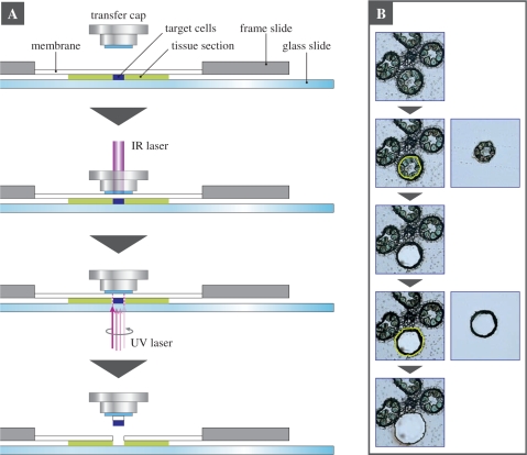
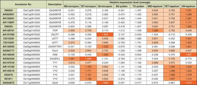
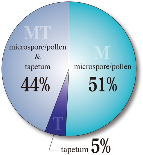
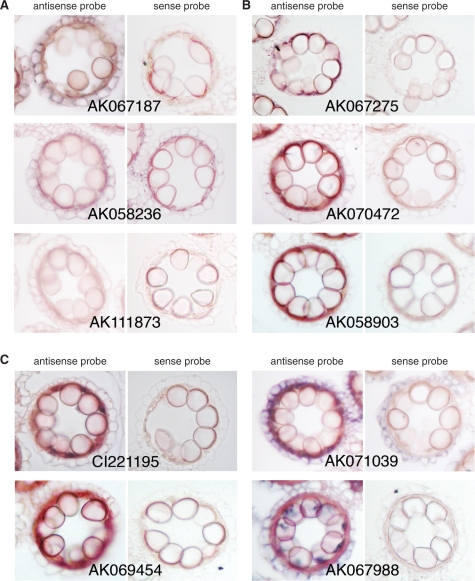
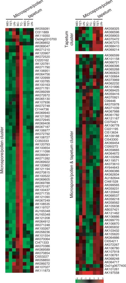
In flowering plants, the male gametophyte, the pollen, develops in the anther. Complex patterns of gene expression in both the gametophytic and sporophytic tissues of the anther regulate this process. The gene expression profiles of the microspore/pollen and the sporophytic tapetum are of particular interest. In this study, a microarray technique combined with laser microdissection (44K LM-microarray) was developed and used to characterize separately the transcriptomes of the microspore/pollen and tapetum in rice. Expression profiles of 11 known tapetum specific-genes were consistent with previous reports. Based on their spatial and temporal expression patterns, 140 genes which had been previously defined as anther specific were further classified as male gametophyte specific (71 genes, 51%), tapetum-specific (seven genes, 5%) or expressed in both male gametophyte and tapetum (62 genes, 44%). These results indicate that the 44K LM-microarray is a reliable tool to analyze the gene expression profiles of two important cell types in the anther, the microspore/pollen and tapetum.


Figures

Fig. 1
Fig. 1
Scheme of a cross-section of a rice anther containing immature microspores. L, locule; T, tapetum; ML, middle layer; En, endothecium; E, epidermis; M, microspores; V, vascular bundle.
Fig. 2
Fig. 2
Laser microdissection (LM) of pollen/microspores and tapetum cells from a cross-section of a rice anther. (A) A schematic procedure for isolating plant target cells by laser microdissection using the Veritas Laser Microdissection System (Molecular Devices), which can use both laser capture and laser cutting. In brief, a transfer cap is placed on a tissue section, which contains the target cells, mounted on a membrane of a frame slide. To anchor and capture the target cells to the transfer cap, a focused low energy infrared (IR) laser beam is targeted on the cells of interest through the transfer cap, causing the cap to fuse to the target cells. The area around the target cells is cut by a UV laser beam. The transfer cap is removed from the tissue section. The laser-microdissected target cells that fused to the transfer cap are separated from the rest of the tissue. (B) Isolation of the pollen/microspore and tapetum cells by the two-step LM. In the case of the TET anther, the microspores were isolated by the first cutting, and the tapetum cells were isolated by the second cutting.
Fig. 3
Fig. 3
Gene expression profiles of reported genes in the 44K LM-microarray. Expression level of 19 probes corresponding to 11 selected genes, which were characterized by previous reports. The same gene accession numbers were annotated to independent probes in the case of OsGAMYB (X98355) and UDT1 (CI515529). Relative values of 0.5–1.0, 1.0–1.5 and 1.5–2.0 were labeled by light orange, orange and dark orange, respectively.
Fig. 4
Fig. 4
Classification of 140 anther-specific genes from the previous 4K microarray in the 44K LM-microarray. Expression profiles of the 44K LM-microarray for the genes reported by Endo et al. (2004) were classified into three spatial expression patterns; microspore/pollen-specific, tapetum-specific and expressed in both tissues.
Fig. 5
Fig. 5
Localization of the anther-specific transcripts in rice anthers. Representative results of RNA in situ hybridization of microspore/pollen-specific genes (A), tapetum-specific genes (B) and genes expressed in both microspore/pollen and tapetum (C). DIG-labeled antisense and sense (control) RNA probes were hybridized to the cross-sections of rice anthers containing immature microspores.
Fig. 6
Fig. 6
Heat map view of the previously reported anther-specific genes differentially expressed in five stages of pollen/microspores and three stages of tapetum cells in the 44K LM-microarray. Cluster analysis of expression profiles of the 140 probes corresponding to the anther-specific genes characterized in the 4K array (Endo et al. 2004). Red indicates higher expression while green represents lower expression.

Similar articles

Cited by

References

    1. Amagai M, Ariizumi T, Endo M, Hatakeyama K, Kuwata C, Shibata D, et al. Identification of anther-specific genes in a cruciferous model plant, Arabidopsis thaliana, by using a combination of Arabidopsis macroarray and mRNA derived from Brassica oleracea. Sex. Plant Reprod. 2003;15:213–222.
    1. Ariizumi T, Hatakeyama K, Hinata K, Inatsugi R, Nishida I, Sato S, et al. Disruption of the novel plant protein NEF1 affects lipid accumulation in the plastids of the tapetum and exine formation of pollen, resulting in male sterility in Arabidopsis thaliana. Plant J. 2004;39:170–1814. - PubMed
    1. Asano T, Masumura T, Kusano H, Kikuchi S, Kurita A, Shimada H, et al. Construction of a specialized cDNA library from plant cells isolated by laser capture microdissection: toward comprehensive analysis of the genes expressed in the rice phloem. Plant J. 2002;32:401–408. - PubMed
    1. Becker JD, Boavida LC, Carneiro J, Haury M, Feijó JA. Transcriptional profiling of Arabidopsis tissues reveals the unique characteristics of the pollen transcriptome. Plant Physiol. 2003;133:713–725. - PMC - PubMed
    1. Cai S, Lashbrook CC. Laser capture microdissection of plant cells from tape-transferred paraffin sections promotes recovery of structurally intact RNA for global gene profiling. Plant J. 2006;48:628–637. - PubMed

Publication types