. 2008 Dec;149(12):5984-95.
doi: 10.1210/en.2008-0680. Epub 2008 Aug 28.

Modulation of Runx2 activity by estrogen receptor-alpha: implications for osteoporosis and breast cancer

Affiliations

Modulation of Runx2 activity by estrogen receptor-alpha: implications for osteoporosis and breast cancer

Omar Khalid et al. Endocrinology. 2008 Dec.

Abstract

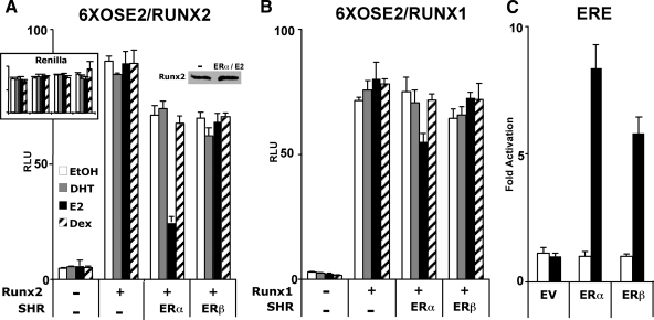
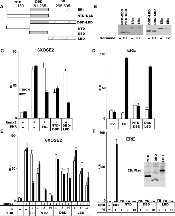
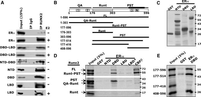
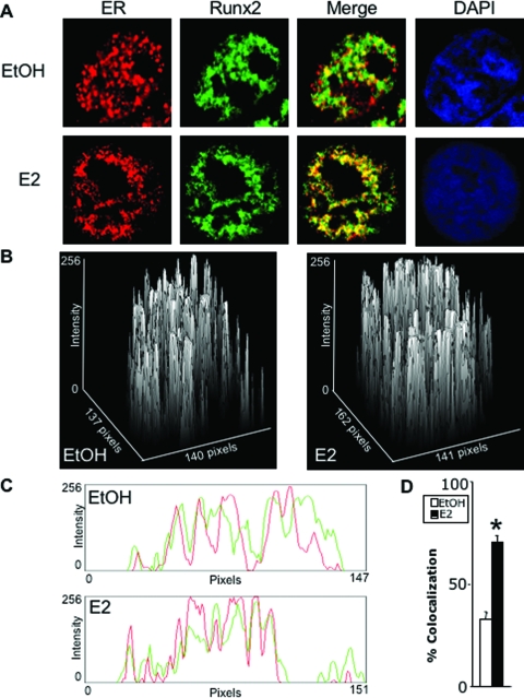
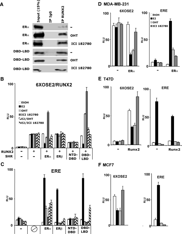
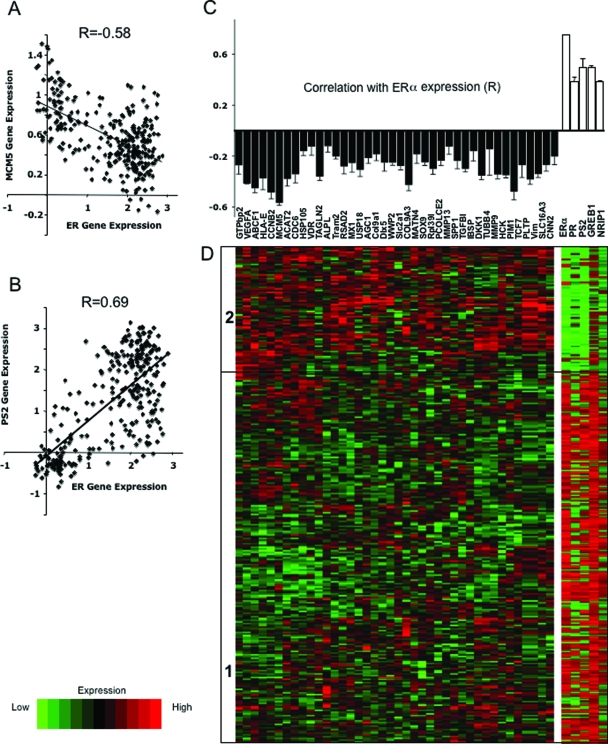
The transcription factors Runx2 and estrogen receptor-alpha (ERalpha) are involved in numerous normal and disease processes, including postmenopausal osteoporosis and breast cancer. Using indirect immunofluorescence microscopy and pull-down techniques, we found them to colocalize and form complexes in a ligand-dependent manner. Estradiol-bound ERalpha strongly interacted with Runx2 directly through its DNA-binding domain and only indirectly through its N-terminal and ligand-binding domains. Runx2's amino acids 417-514, encompassing activation domain 3 and the nuclear matrix targeting sequence, were sufficient for interaction with ERalpha's DNA-binding domain. As a consequence of the interaction, Runx2's transcriptional activation activity was strongly repressed, as shown by reporter assays in COS7 cells, breast cancer cells, and late-stage MC3T3-E1 osteoblast cultures. Metaanalysis of gene expression in 779 breast cancer biopsies indicated negative correlation between the expression of ERalpha and Runx2 target genes. Selective ER modulators (SERM) induced ERalpha-Runx2 interactions but led to various functional outcomes. The regulation of Runx2 by ERalpha may play key roles in osteoblast and breast epithelial cell growth and differentiation; hence, modulation of Runx2 by native and synthetic ERalpha ligands offers new avenues in selective ER modulator evaluation and development.


Figures

Figure 1
Figure 1
ERα inhibits Runx2. A and B, COS7 cells were transiently transfected with the Runx reporter 6XOSE2-luc (firefly) along with expression vectors encoding human Runx2 (A), Runx1 (B), ERα, and/or ERβ as indicated. Cells were treated with dihydrotestosterone (DHT), (E2) Estradiol, dexamethasone (Dex) (10 nm each), or ethanol (EtOH) vehicle (0.01%) for 24 h and subjected to luciferase assay as described in Materials and Methods. C, COS7 cells were transiently transfected with an ER reporter (ERE-luc) along with an expression vector encoding either ERα or ERβ and treated for 24 h with E2, followed by the luciferase assay. In each experiment, the firefly luciferase results were corrected for the expression of a cotransfected CMV-renilla luciferase construct (internal control), except for A, where the renilla luciferase values are shown in the inset. The immunoblot in A shows that Runx2 expression was not inhibited in the presence of ERα and its ligand. All data are presented as mean values ± sem, with n = 4 dish replicates of a representative experiment, repeated at least three times. EV, Empty vector; RLU, relative light units; SHR, steroid hormone receptor.
Figure 2
Figure 2
Functional mapping of ERα substructures and the dissociation between ERα-mediated transcriptional activation and Runx2 repression. A, Schematic illustration showing full-length and ERα fragments that were transiently expressed in COS7 cells. The NTD, DBD, and LBD fragments were FLAG-tagged to facilitate immunoblot detection shown in the inset of F. B, Immunoblot analysis of cells transfected with the indicated constructs using antibodies against ERα NTD (sc-7207, left blot) or ERα LBD (sc-787, right blot). C–F, COS7 cells were transiently transfected with either the 6XOSE2-luc (C and E) or the ERE-luc reporter (D and F), along with the indicated expression plasmids. Luciferase activity was measured 24 h after treatment with either ethanol (0.01%) or E2 (10 nm). All data are presented as mean values ± sem, with n = 4 dish replicates of a representative experiment, repeated at least three times.
Figure 3
Figure 3
Interaction between ERα and Runx2 domains in Co-IP and GST pull-down assays. A, COS7 cells were transiently transfected with plasmids encoding each of the specified ERα fragments and Runx2, followed by 24 h treatment with either 10 nm E2 or vehicle. The ERα and its fragments were detected in either whole-cell extracts as input or IgG or Runx2 immunoprecipitates. B, Schematic diagram of full-length (FL) Runx2 and fragments transcribed and translated in vitro. The scheme at the top depicts the three Runx2 domains. Boxes with 1, 2, 3, or N mark the positions of the respective activation domains and the nuclear matrix targeting sequences. The thick line above the PST domain represents the surface interacting with ERα. C, Coomassie-stained SDS-PAGE of the bacterially expressed and purified GST or GST fusion proteins used as baits in the pull-down assays. D and E, A mixture of the indicated radiolabeled Runx2 fragments was incubated with the depicted GST-fusion proteins used as baits. The positive control GST-CBFβ was used as a bait for the Runt domain. The autoradiograph shows the fragments pulled down by the indicated baits. Asterisk in D denotes presence of 10 nm E2.
Figure 4
Figure 4
Immunofluorescence of ERα and Runx2. COS7 cells were transiently transfected with ERα and Runx2 and treated for 24 h with ethanol vehicle (EtOH) or E2 (10 nm). A–C, ERα (red) and Runx2 (green) were visualized using confocal microscopy as described in Materials and Methods. Colocalization (yellow in A) is demonstrated by surface plots (B) and by red/green profiles (C). D, Ten cells were randomly selected from each of a set of four untreated and four treated cultures, and colocalization was quantified and plotted as mean ± sem. *, P = 8.3 × 10⁻⁸. DAPI, 4′,6-Diamidino-2-indole.
Figure 5
Figure 5
Developmental stage-specific inhibition of Runx2 by E2 in osteoblasts. A, MC3T3-E1 cells were treated with ethanol or E2 (10 nm) and the presence of ERα in Runx2 immunocomplexes was examined as described in Fig. 3A. B–E, MC3T3-E1 cells stably transfected with the 6XOSE2-luc Runx2 reporter were subjected to differentiation conditions and treated with ethanol (white bars) or E2 (10 nm; black bars) commencing at confluence. Levels of the indicated mRNAs were measured on d 4, 11, and 18 by quantitative RT-PCR as described in Materials and Methods. Data were corrected for the expression of ribosomal protein L10A, which itself did not significantly change during culture progression or in response to E2. RANKL was not expressed on d 4, and the low expression of OC and luciferase on this day are shown in the respective insets (mean ± sd; n = 3).
Figure 6
Figure 6
Various effects of SERM-bound ERα on Runx2. A, Co-IP assays were performed as in Fig. 3A after treatment of COS7 cells with 100 nm OHT or 100 nm ICI 182780. B and C, COS7 cells were transfected with Runx2 and its 6XOSE2-luc reporter (B) or with ERE-luc (C) along with expression vectors coding for the indicated ER isoforms or fragments. Cells were treated for 24 h with ethanol, E2 (10 nm), OHT (100 nm), ICI 182780 (100 nm), or combinations thereof and then subjected to luciferase assays. D–F, Three breast cancer cell lines, MDA-MB-231 (D), T47D (E), and MCF7 (F), were transfected with the 6XOSE2-luc (left) or the ERE-luc (right) reporter, along with expression vectors for ERα (D), Runx2 (E), or empty vector control and treated with 10 nm E2, 100 nm OHT, or 100 nm ICI 182780 as indicated. All data are presented as mean values ± sem, with n = 4 dish replicates of a representative experiment, repeated at least three times.
Figure 7
Figure 7
Metaanalysis of the correlation between expression of ERα and Runx2 target genes in breast cancer biopsies. A and B, Scatter plots of the expression of MCM5 (A) or pS2 (B) vs. ERα in 286 beast cancer biopsies previously subjected to comprehensive gene expression analysis (39). C, Correlation coefficients between the expression of ERα and each of 40 Runx2 target genes based on metaanalysis of 779 breast cancer biopsies described in three published databases (42,43,44). Each of the correlation coefficients was significant with P < 10−4. D, Expression levels of the Runx2 target genes in one cohort of 286 breast cancer biopsies (39) was subjected to an unsupervised cluster analysis, resulting in two major branches of tumor samples designated in the heat map as 1 and 2. The expression levels of ERα and four ERα target genes in each of the 286 biopsies are represented as a heat map on the right.

References

    1. Coffman JA 2003 Runx transcription factors and the developmental balance between cell proliferation and differentiation. Cell Biol Int 27:315–324 - PubMed
    1. Schroeder TM, Jensen ED, Westendorf JJ 2005 Runx2: a master organizer of gene transcription in developing and maturing osteoblasts. Birth Defects Res C Embryo Today 75:213–225 - PubMed
    1. Komori T, Yagi H, Nomura S, Yamaguchi A, Sasaki K, Deguchi K, Shimizu Y, Bronson RT, Gao YH, Inada M, Sato M, Okamoto R, Kitamura Y, Yoshiki S, Kishimoto T 1997 Targeted disruption of Cbfa1 results in a complete lack of bone formation owing to maturational arrest of osteoblasts. Cell 89:755–764 - PubMed
    1. Otto F, Thornell AP, Crompton T, Denzel A, Gilmour KC, Rosewell IR, Stamp GW, Beddington RS, Mundlos S, Olsen BR, Selby PB, Owen MJ 1997 Cbfa1, a candidate gene for cleidocranial dysplasia syndrome, is essential for osteoblast differentiation and bone development. Cell 89:765–771 - PubMed
    1. Salingcarnboriboon R, Tsuji K, Komori T, Nakashima K, Ezura Y, Noda M 2006 Runx2 is a target of mechanical unloading to alter osteoblastic activity and bone formation in vivo. Endocrinology 147:2296–2305 - PubMed

Publication types

MeSH terms