. 2008 Sep 2;105(35):13087-92.
doi: 10.1073/pnas.0802220105. Epub 2008 Aug 28.

Generation of functional erythrocytes from human embryonic stem cell-derived definitive hematopoiesis

Affiliations

Generation of functional erythrocytes from human embryonic stem cell-derived definitive hematopoiesis

Feng Ma et al. Proc Natl Acad Sci U S A. .

Abstract

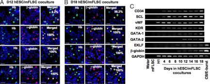
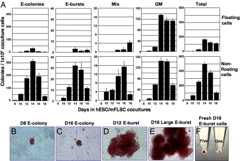
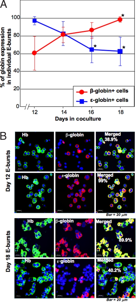
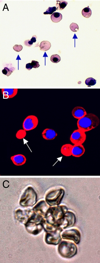
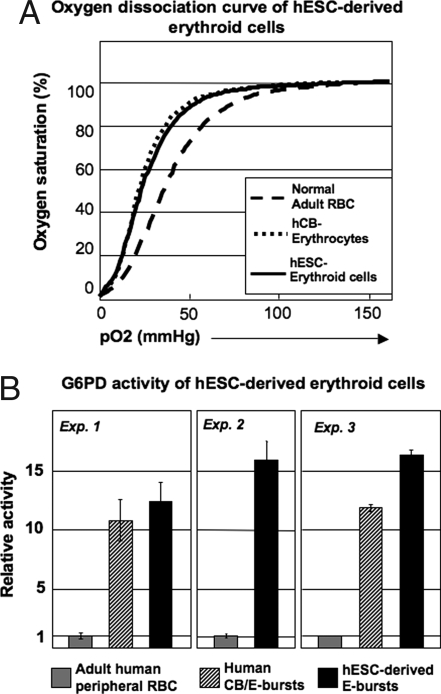
A critical issue for clinical utilization of human ES cells (hESCs) is whether they can generate terminally mature progenies with normal function. We recently developed a method for efficient production of hematopoietic progenitors from hESCs by coculture with murine fetal liver-derived stromal cells. Large numbers of hESCs-derived erythroid progenitors generated by the coculture enabled us to analyze the development of erythropoiesis at a clone level and investigate their function. The results showed that the globin expression in the erythroid cells in individual clones changed in a time-dependent manner. In particular, embryonic epsilon-globin-expressing erythroid cells from individual clones decreased, whereas adult-type beta-globin-expressing cells increased to approximately 100% in all clones we examined, indicating that the cells undergo definitive hematopoiesis. Enucleated erythrocytes also appeared among the clonal progeny. A comparison analysis showed that hESC-derived erythroid cells took a similar differentiation pathway to human cord blood CD34(+) progenitor-derived cells when examined for the expression of glycophorin A, CD71 and CD81. Furthermore, these hESC-derived erythroid cells could function as oxygen carriers and had a sufficient glucose-6-phosphate dehydrogenase activity. The present study should provide an experimental model for exploring early development of human erythropoiesis and hemoglobin switching and may help in the discovery of drugs for hereditary diseases in erythrocyte development.


Conflict of interest statement

The authors declare no conflict of interest.

Figures

Fig. 1.
Fig. 1.
Time-associated changes in expression of globins and hematopoiesis-related genes in hESC/mFLSC-derived erythroid cells. (A) Immunostaining of Hb, β-, ε-, α-, and γ-globins in cells from day-12 hESC/mFLSC cocultures. β-Globin was expressed only in 8.2% of the Hb+ erythroid cells, whereas ε-, α-, and γ-globins were expressed in 100% of Hb+ erythroid cells. (Scale bar: 25 μm.) (B) Immunostaining of Hb, β-, ε-, α-, and γ-globins in cells from day-18 hESC/mFLSC coculture cells. β-Globin+ cells increased to 56.2% of Hb+ erythroid cells, although ε-, α-, and γ-globins were still expressed in all Hb+ erythroid cells. (Scale bar: 25 μm.) (C) Time course of expression of early hematopoiesis-related genes and the definitive hematopoiesis β-globin gene during the hESC/mFLSC coculture detected by RT-PCR.
Fig. 2.
Fig. 2.
Generation of erythroid progenitors in hESC/mFLSC coculture. (A) Generation of CFCs from floating and nonfloating cells over time in hESC/mFLSC cocultures. (B–E) Micrographs of E colonies derived from day-8 (B) and day-16 (C) cocultures and E bursts from day-12 (D) and day-16 (E) cocultures. (F) Photo of harvested E burst cells from day-16 coculture, showing the red color of human erythroid cells. A total of 2 × 10⁵ (Right) and 1 × 10⁶ (Left) erythroid cells were collected from one and five E-bursts, respectively.
Fig. 3.
Fig. 3.
Clonal analysis of time-associated changes in globin expression during hESC/mFLSC coculture. (A) Globin expression in E bursts prepared after different amounts of time in coculture (n = 6 in each time point). Each E burst was individually picked from colony culture medium, and the globin expression was examined by immunostaining. The percentages were calculated from the ratio of β- or ε-globin+/human Hb+ cells. *, P < 0.01 when compared with the average expression of β- and ε-globins in day-12 cocultures. (B) Representative micrographs of Hb and β- and ε-globin immunostaining of E burst cells derived from E-BFCs at days 12 and 18 of coculture. (Scale bar: 20 μm.)
Fig. 4.
Fig. 4.
Clonal analysis of progressive maturation of hESC/mFLSC coculture-derived erythroid cells. (A) Cytospin sample of hESC-derived erythroid cells from a day-12+6 suspension culture (May-Grunwald-Giemsa staining). Arrows indicate enucleated erythrocytes. (B) Immunostaining for β-globin expression in hESC-derived erythroid cells from the same suspension culture shown in A. Arrows indicate β-globin-expressing enucleated erythrocytes. (C) Cluster of enucleated erythrocytes derived from hESCs from the same suspension culture shown in A and B.
Fig. 5.
Fig. 5.
Functional assays of hESC-derived erythroid cells. (A) Oxygen dissociation curves of day-15 hESC/mFLSC coculture-derived clonal E burst erythroid cells at day 16 of colony culture, human CB, and adult PB. (B) G6PD activity of day-15 hESC/mFLSC coculture-derived clonal E burst erythroid cells at day 16 of the colony culture compared with human CB-derived E burst cells and adult PB.

References

    1. Orkin S-H, Zon L-I. Hematopoesis and stem cells: Plasticity versus developmental heterogeneity. Nat Immunol. 2002;3:323–328. - PubMed
    1. Palis J, Segel G-B. Developmental biology of erythropoiesis. Blood Rev. 1998;12:106–114. - PubMed
    1. Thomson J-A, et al. Embryonic stem cell lines derived from human blastocysts. Science. 1998;282:1145–1147. - PubMed
    1. Kaufman D-S, Hanson E-T, Lewis R-L, Auerbach R, Thomson J-A. Hematopoietic colony-forming cells derived from human embryonic stem cells. Proc Natl Acad Sci USA. 2001;98:10716–10721. - PMC - PubMed
    1. Wang L-S, et al. Endothelial and hematopoietic cell fate of human embryonic stem cells originates from primitive endothelium with hemangioblastic properties. Immunity. 2004;21:31–41. - PubMed

Publication types

MeSH terms

LinkOut - more resources