. 2008 Sep 9;105(36):13421-6.
doi: 10.1073/pnas.0801613105. Epub 2008 Aug 28.

miR-34a repression of SIRT1 regulates apoptosis

Affiliations

miR-34a repression of SIRT1 regulates apoptosis

Munekazu Yamakuchi et al. Proc Natl Acad Sci U S A. .

Abstract

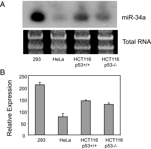
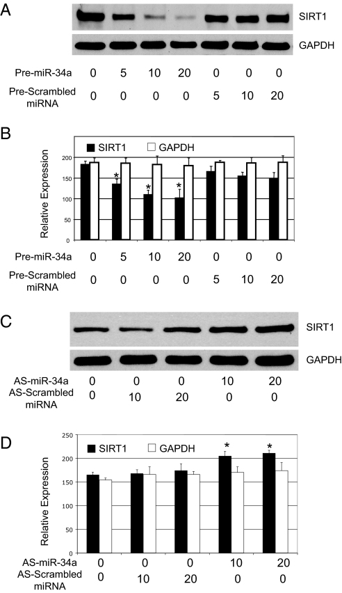
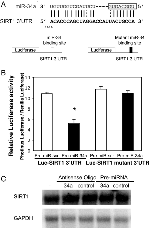
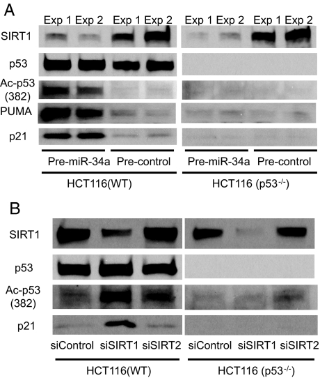
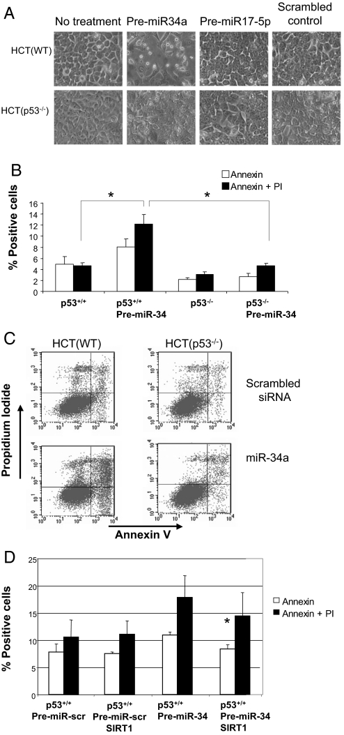
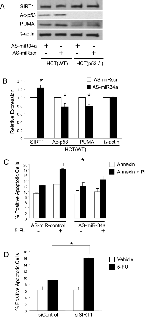
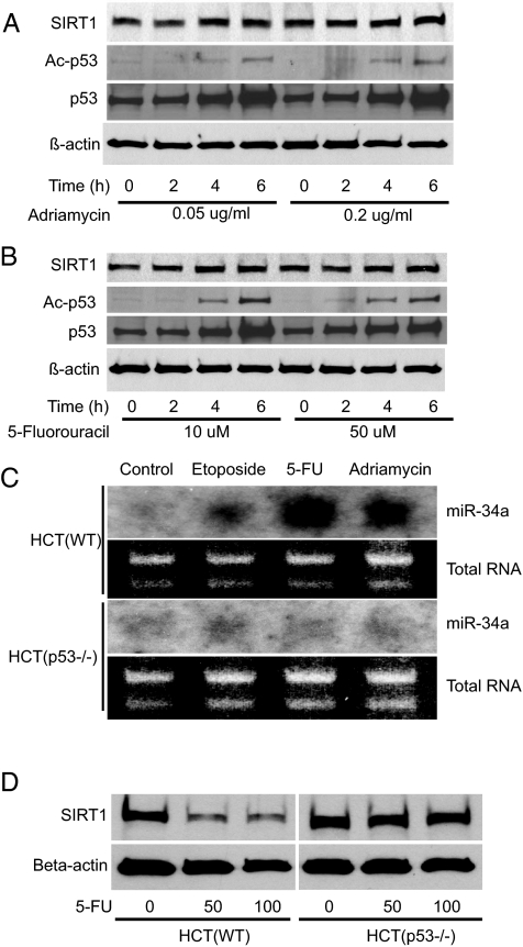
MicroRNA 34a (miR-34a) is a tumor suppressor gene, but how it regulates cell proliferation is not completely understood. We now show that the microRNA miR-34a regulates silent information regulator 1 (SIRT1) expression. MiR-34a inhibits SIRT1 expression through a miR-34a-binding site within the 3' UTR of SIRT1. MiR-34 inhibition of SIRT1 leads to an increase in acetylated p53 and expression of p21 and PUMA, transcriptional targets of p53 that regulate the cell cycle and apoptosis, respectively. Furthermore, miR-34 suppression of SIRT1 ultimately leads to apoptosis in WT human colon cancer cells but not in human colon cancer cells lacking p53. Finally, miR-34a itself is a transcriptional target of p53, suggesting a positive feedback loop between p53 and miR-34a. Thus, miR-34a functions as a tumor suppressor, in part, through a SIRT1-p53 pathway.


Conflict of interest statement

The authors declare no conflict of interest.

Figures

Fig. 1.
Fig. 1.
Expression of miR-34a. (A) Human cell lines, including human colon cancer (HCT116) cells by Northern blot (repeated three times with similar results). (B) Densitometric quantification of Northern analysis of miR-34a repeated three times. Both p53+/+ and p53−/− HCT116 cells have similar amounts of miR-34a, in the absence of genotoxic damage.
Fig. 2.
Fig. 2.
SIRT1 is a target of microRNA miR-34. (A) Dose-dependent suppression of SIRT1 by miR-34a in HeLa cells. HeLa cells were transfected with 5, 10, or 20 nM miRNA precursors and immunoblotted as above. (B) Quantification by densitometry of effects of premiR-34a on SIRT1 expression (n = 3 ± SD; *, P < 0.05 compared with scrambled mRNA). (C) Dose-dependent increase of SIRT1 by miR-34a in HEK293 cells. HEK293 cells were transfected with 15 or 30 nM antisense miRNA and immunoblotted as above. (D) Quantification by densitometry of effects of knocking down endogenous miR-34a on SIRT1 expression (n = 3 ± SD; *, P < 0.05 compared with scrambled mRNA).
Fig. 3.
Fig. 3.
MiR-34a binding site within SIRT1 3′ UTR mediates miR-34a translational repression. (A Top) miR-34a and the miR-34a-binding site in the 3′ UTR of SIRT1. (Bottom) Design of a miR-34a reporter vector containing a CMV promoter driving expression of a luciferase cDNA fused to the SIRT1 3′ UTR or to a mutated SIRT1 3′ UTR. (B) The 3′ UTR of SIRT1 mediates miR-34 control of SIRT1 expression. WT HCT116 cells were transfected with a reporter vector consisting of a luciferase cDNA fused to the 3′ UTR of SIRT1 which contains a binding site of miR-34. Another vector contained the luciferase cDNA fused to a SIRT1 3′ UTR with a mutant miR-34a-binding site. The cells were also transfected with a CMV-Renilla luciferase vector as an internal standard. PremiR-34a decreases expression of luciferase containing a WT miR-34a binding site (Left) but not a mutant binding site (Right). (C) miR-34 does not change SIRT1 mRNA. HCT116 cells were transfected with antisense-miR-34a or PremiR-34a, and total RNA was harvested and analyzed for SIRT1 by Northern blotting.
Fig. 4.
Fig. 4.
miR-34a increases acetylation of p53 by decreasing SIRT1 expression. (A) miR-34 decreases SIRT1, increases acetylated p53, and increases two transcriptional targets of p53. In duplicate experiments, HCT(WT) and HCT(p53−/−) were transfected with precursors to microRNA and incubated for 36 h. SIRT1, acetylated p53, PUMA, and p21 were measured by immunoblotting. These experiments were performed in duplicate. (B) SIRT1 regulates acetylation of p53. HCT(WT) and HCT(p53−/−) were transfected with siRNA for SIRT1 or SIRT2, incubated for 36 h, and lysates were immunoblotted. Knockdown of SIRT1 permits an increase in p53 acetylation and an increase in p21, a transcriptional target of p53.
Fig. 5.
Fig. 5.
miR-34 activates apoptosis. (A) p53 mediates miR-34 induced apoptosis. HCT(WT) and HCT(p53−/−) cells were transfected with premiR-34 or control and then photographed. Overexpression of miR-34a causes cells to detach and to round up but only in cells expressing p53. (B) HCT(WT) and HCT(p53−/−) cells were treated as above, and apoptosis was measured by Annexin V and propidium iodine staining. (C) HCT(WT) and HCT(p53−/−) cells were treated as above, and apoptosis was measured by FACS (n = 3 ± SD; *, P = 0.003). Overexpression of miR-34a induces apoptosis but only in the presence of p53. (D) SIRT1 rescues cells from apoptosis after overexpression of miR-34. HCT(WT) cells were transfected with premiR-34 or control and with a vector expressing SIRT1 or control, and apoptosis was measured by FACS (n = 3 ± SD; *, P = 0.01 vs. PremiR-34 without SIRT1). Overexpression of SIRT1 blocks apoptosis induced by miR-34a.
Fig. 6.
Fig. 6.
Endogenous miR-34a regulates SIRT1. (A) HCT cells were transfected with antisense oligonucleotides to miR-34 (antisense-miR-34a) or scrambled oligonucleotides. Cells were treated with 5-FU for 16 h to activate p53, and cell lysates were immunoblotted for SIRT1, acetylated p53, and the p53 regulated gene product PUMA. Knockdown of endogenous miR-34a increases SIRT1, decreases acetylation of p53, and decreases protein levels of PUMA. (B) Quantitative effects of knockdown of endogenous miR-34a on Ac-p53. The above immunoblots were repeated three times and quantified by densitometry (n = 3 ± SD; *, P < 0.05 vs. scrambled miRNA). (C) Genotoxic stress increases apoptosis in HCT(WT) cells, and miR-34a mediates part of the genotoxic effect. HCT(WT) cells were transfected with antisense-miR-34a or control oligonucleotides, exposed to 5-FU, and apoptosis was measured as above. Knockdown of miR-34a decreases apoptosis (n = 3 ± SD; *, P < 0.05). (D) Knockdown of SIRT1 increases apoptosis. HCT(WT) cells were transfected with siControl or siSIRT1 then treated with vehicle or control. Annexin V staining was measured by FACS (n = 3 ± SD; *, P < 0.005).
Fig. 7.
Fig. 7.
p53 regulates miR-24a expression. HCT cells were treated with genotoxic agents, and cell lysates were assayed for p53, SIRT1, and miR-34a. Acetylation of p53 is increased after treatment with (A) adriamycin and (B) 5-FU. (C) Genotoxic agents increase miR-34a only in HCT cells expressing p53. (D) Genotoxic agents lead to a decrease in SIRT1 levels but only in cells expressing p53.

References

    1. Fong CT, et al. Loss of heterozygosity for the short arm of chromosome 1 in human neuroblastomas: Correlation with N-myc amplification. Proc Natl Acad Sci USA. 1989;86:3753–3757. - PMC - PubMed
    1. Rigaud G, et al. Allelotype of pancreatic acinar cell carcinoma. Int J Cancer. 2000;88:772–777. - PubMed
    1. Bello MJ, et al. Allelic status of chromosome 1 in neoplasms of the nervous system. Cancer Genet Cytogenet. 1995;83:160–164. - PubMed
    1. Moley JF, et al. Consistent association of 1p loss of heterozygosity with pheochromocytomas from patients with multiple endocrine neoplasia type 2 syndromes. Cancer Res. 1992;52:770–774. - PubMed
    1. Genuardi M, Tsihira H, Anderson DE, Saunders GF. Distal deletion of chromosome Ip in ductal carcinoma of the breast. Am J Hum Genet. 1989;45:73–82. - PMC - PubMed

Publication types