. 2008 Sep 26;321(5897):1841-4.
doi: 10.1126/science.1161621. Epub 2008 Aug 28.

Seeding and propagation of untransformed mouse mammary cells in the lung

Affiliations

Seeding and propagation of untransformed mouse mammary cells in the lung

Katrina Podsypanina et al. Science. .

Abstract

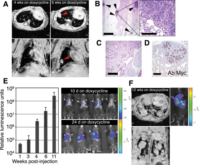
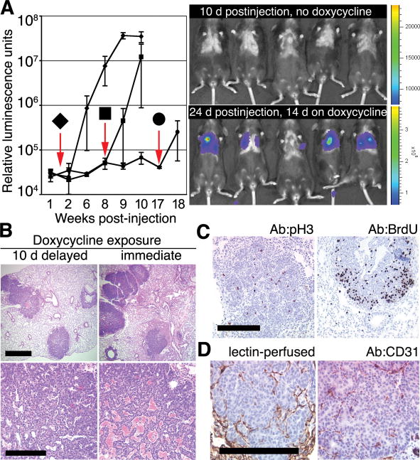
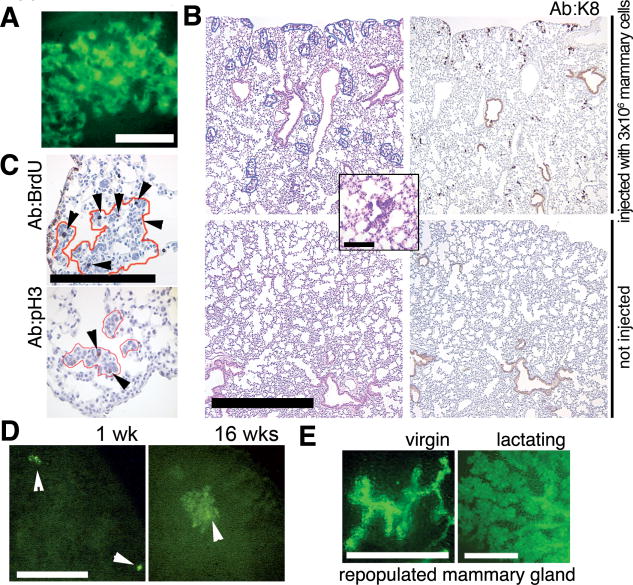
The acquisition of metastatic ability by tumor cells is considered a late event in the evolution of malignant tumors. We report that untransformed mouse mammary cells that have been engineered to express the inducible oncogenic transgenes MYC and Kras(D12), or polyoma middle T, and introduced into the systemic circulation of a mouse can bypass transformation at the primary site and develop into metastatic pulmonary lesions upon immediate or delayed oncogene induction. Therefore, previously untransformed mammary cells may establish residence in the lung once they have entered the bloodstream and may assume malignant growth upon oncogene activation. Mammary cells lacking oncogenic transgenes displayed a similar capacity for long-term residence in the lungs but did not form ectopic tumors.


Figures

Fig. 1
Fig. 1
Untransformed mouse mammary cells form lung metastases after IV injection and induction of oncogenes. (A to D) Lung metastases develop from intravenously injected phenotypically normal mammary cells upon activation of MYC and KrasD12 transgenes. (A) Metastases were monitored by MRI in Rag1−/− mice after IV delivery of 1 × 10⁶ dissociated primary mammary cells from doxycycline-naïve TOM;TOR;MTB mice. Recipient mice were fed doxycycline for 6 weeks starting 1 day before injection. Representative axial (top) and coronal (bottom) images obtained from the same animal 4 and 6 weeks after injection show development of a solid nodule (arrowheads) in the lung. (B) Foci of hematoxylin-eosin (H/E)–stained mammary adenocarcinoma (arrowheads) in paraffin-embedded lung sections of the same Rag1−/− mouse as in (A). Scale bars indicate 1 mm (left) and 0.1 mm (right). (C) No tumors were observed in lung sections of Rag1−/− mice that did not receive doxycycline after IV delivery of 1 × 10⁶ primary mammary cells from doxycycline-naïve TOM;TOR;MTB mice. Scale bar, 1 mm. (D) Tumor cells from the same animal as in (A), but not the surrounding lung tissue, stained with anti-MYC antisera. Scale bar, 0.1 mm. (E and F) Lung metastases develop from intravenously injected phenotypically normal mammary cells upon activation of a PyMT transgene. (E) Donor cells expressing their transgene were detected by bioluminescence imaging after 5 × 10⁵ primary mammary cells from doxycycline-naïve TOMT:IRES:Luc;MTB mice were injected intravenously into Rag1−/− mice that were placed on doxycycline 1 day before injection. Representative images at day 10 and day 24 after injection show the presence of signal-emitting cells in the thorax (right); temporal increases in bioluminescence (14) were quantified in relative luminescence units (left; n = 5 mice; error bars represent SD). (F) Axial (top) and coronal (bottom) MRI images of a mouse from (E) maintained on doxycycline for 12 weeks show solid nodules in the lung. A corresponding bioluminescence image is shown on the right.
Fig. 2
Fig. 2
Delay in oncogene activation does not preclude the development of ectopic mammary tumors. (A) Bioluminescence in Rag1−/− mice after IV delivery of 1 × 10⁵ mammary cells from doxycycline-naïve TOMT:IRES:Luc;MTB mice is undetectable before doxycycline exposure but can be induced at various times after placing mice on doxycycline 1.5 (◆, n = 7 mice), 8 (■, n = 3 mice), or 17 weeks (●, n = 2 mice) after IV injection. Downward arrows indicate times of addition of doxycycline to the diet. Error bars represent SD. Representative bioluminescence images (right) obtained 10 days after injection in the absence of doxycycline (top right) and after 2 additional weeks on doxycycline (bottom right). (B) Histologically similar metastatic tumors in lungs of Rag1−/− mice after IV delivery of 1 × 10⁵ mammary cells from doxycycline-naïve TOMT:IRES:Luc;MTB mice exposed to doxycycline for 8 weeks starting 10 days after (left) or 1 day before (right) IV injection. Scale bars, 1 mm (top) and 0.1 mm (bottom). (C) Mitotic activity in tumor foci in lung sections from Rag1−/− recipient of TOM;TOR;MTB cells (left; stained with anti-pH3) or from Rag1−/− recipient of TOMT:IRES:Luc;MTB cells (right; stained with anti-BrdU serum after BrdU labeling) (14). Scale bar, 0.1 mm. (D) Angiogenic proficiency demonstrated by perfusion of the ectopic tumor with biotinylated lectin (left) (14) and by staining endothelial cells within tumor foci with anti-CD31 serum in a Rag−/− recipient of the TOMT:IRES:Luc; MTB cells (right). Scale bar, 0.1 mm.
Fig. 3
Fig. 3
Mammary cells without an oncogenic transgene can persist in the lung. (A) Focus of green cells observed under excitation light (14) in a whole fresh lung of a Rag1−/− recipient 3 weeks after IV injection of 5 × 10⁵ dissociated mammary cells from a β-actin-GFP mouse. Scale bar, 0.1 mm. (B) Representative size and distribution of the H/E–stained ectopic foci in lung sections from Rag1−/− mice injected intravenously with 3 × 10⁶ mammary cells from a β-actin-GFP donor at 3 weeks after injection (top left). Inset shows a representative H/E–stained focus at high magnification. A consecutive section (top right) was stained with rat antiserum against K8. No foci were detected by H/E (bottom left) or K8 staining (bottom right) of lung sections from uninjected Rag1−/− mice. Scale bar, 1 mm. Inset scale bar, 0.2 mm. (C) Mitotic activity in ectopic epithelial outgrowths (outlined in red) demonstrated by BrdU-labeling detected with rat anti-BrdU serum (arrowheads, top) or by staining with anti-pH3 (arrowheads, left). Scale bar, 0.2 mm. (D) Larger foci of green fluorescent cells were observed by whole-lung imaging under excitation light (14) at 16 weeks after injection (right) as compared with 1 week after injection (left). Rag1−/− recipients were injected with 3 × 10⁵ dissociated mammary cells from a β-actin-GFP mouse. Scale bar, 1 mm. (E) Mammary gland repopulation in secondary Rag1−/− recipients produces a green fluorescent mammary tree detectable under excitation light in the whole-mount preparations 4 or 7 weeks after transplantation (14). Glands were harvested from virgin recipients (left), or host animals were mated and the transplanted glands harvested 1 day postpartum (right). Scale bar, 1 mm.

Comment in

  • Cancer. The metastasis cascade.
    Klein CA. Klein CA. Science. 2008 Sep 26;321(5897):1785-7. doi: 10.1126/science.1164853. Science. 2008. PMID: 18818347 No abstract available.

References

    1. Hanahan D, Weinberg RA. Cell. 2000;100:57. - PubMed
    1. Husemann Y, et al. Cancer Cell. 2008;13:58. - PubMed
    1. Gupta GP, et al. Nature. 2007;446:765. - PubMed
    1. Gupta PB, et al. Nat Genet. 2005;37:1047. - PMC - PubMed
    1. Hartwell KA, et al. Proc Natl Acad Sci USA. 2006;103:18969. - PMC - PubMed

Publication types

MeSH terms

Substances