. 2008 Aug 29;321(5893):1206-10.
doi: 10.1126/science.1161302.

Solution structure of the integral human membrane protein VDAC-1 in detergent micelles

Affiliations

Solution structure of the integral human membrane protein VDAC-1 in detergent micelles

Sebastian Hiller et al. Science. .

Abstract

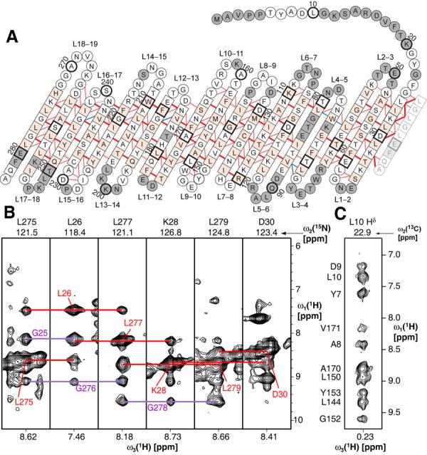
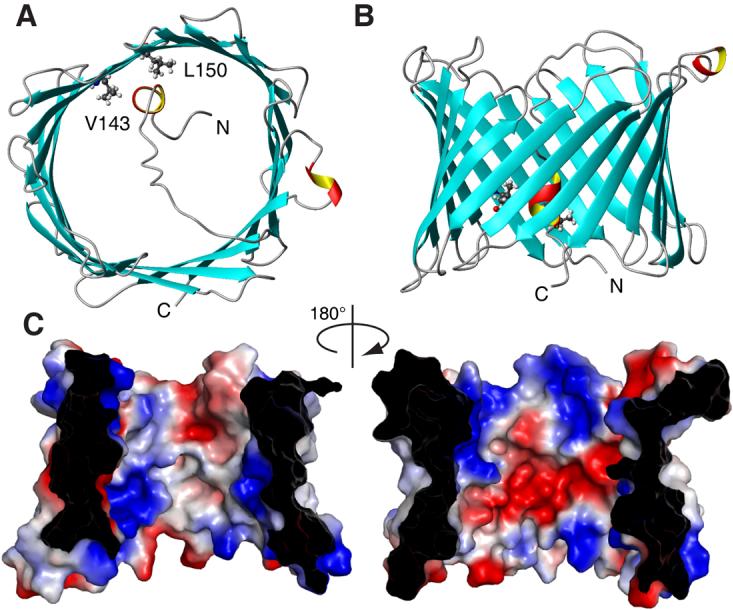
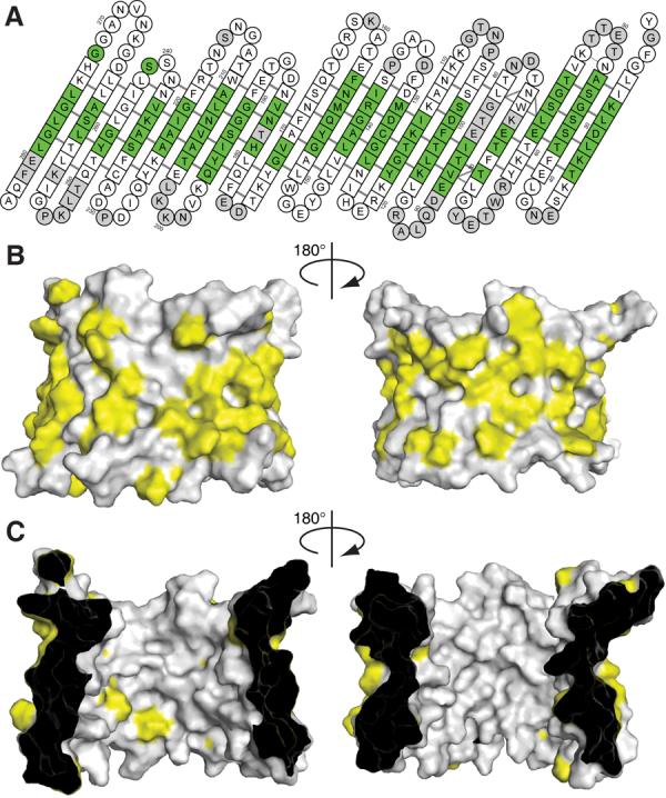
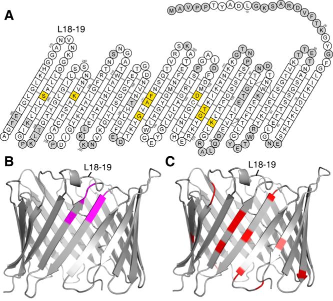
The voltage-dependent anion channel (VDAC) mediates trafficking of small molecules and ions across the eukaryotic outer mitochondrial membrane. VDAC also interacts with antiapoptotic proteins from the Bcl-2 family, and this interaction inhibits release of apoptogenic proteins from the mitochondrion. We present the nuclear magnetic resonance (NMR) solution structure of recombinant human VDAC-1 reconstituted in detergent micelles. It forms a 19-stranded beta barrel with the first and last strand parallel. The hydrophobic outside perimeter of the barrel is covered by detergent molecules in a beltlike fashion. In the presence of cholesterol, recombinant VDAC-1 can form voltage-gated channels in phospholipid bilayers similar to those of the native protein. NMR measurements revealed the binding sites of VDAC-1 for the Bcl-2 protein Bcl-x(L), for reduced beta-nicotinamide adenine dinucleotide, and for cholesterol. Bcl-x(L) interacts with the VDAC barrel laterally at strands 17 and 18.


Figures

Figure 1
Figure 1
Architecture of VDAC-1. (A) The amino acid sequence of VDAC-1 in one-letter code is arranged according to the secondary and tertiary structure. Amino acids in squares denote β-sheet secondary structure as identified by secondary chemical shifts, all other amino acids are in circles. Red and blue lines denote experimentally observed NOE contacts between two amide protons and NOE contacts involving side chain atoms, respectively. Bold lines indicate strong NOEs typically observed between hydrogen bonded residues in β-sheets. For clarity of the presentation, not all observed NOEs are shown. The 19th strand is duplicated at the right next to strand 1 to allow indicating the barrel-closure NOEs. The side chains of white and orange residues point towards the inside and outside of the barrel, respectively. Dashed lines show probable contacts between protons with degenerate 1H chemical shifts. Grey residues could not be assigned so far. Every tenth amino acid is marked with a heavy outline, and corresponding residue numbers are indicated. (B) Strips from a 3D [1H,1H]-NOESY-15N-TROSY defining the barrel closure between parallel strands 1 and 19. Red lines show the interstrand contacts for the depicted residues, whereas the violet lines indicate the NOE contacts for the respective opposite residues. (C) Strip from a 3D [1H,1H]-NOESY-13C-HMQC taken at the position of a methyl group of Leu 10. The assignments of the individual NOE signals are indicated on the left and exemplify the NOEs defining the location of the N-terminal helix in the barrel.
Figure 2
Figure 2
NMR solution structure of VDAC-1 in LDAO micelles. (A) and (B) Top- and side-view, respectively, of the conformer closest to the mean of the conformational ensemble in ribbon representation. β-sheets are shown blue and α-helical secondary structure in red and yellow. N- and C-termini and residues L150 and V143 are indicated. (C) Van der Waals surface of VDAC-1. The surface is colored according to the surface potential, calculated using vacuum electrostatics in the program Pymol. Blue indicates positive charge and red negative charge.
Figure 3
Figure 3
Hydrophobic surface of VDAC-1. (A) Result of a titration with the spin-labelled detergent 16-DSA. Residues with a relaxation enhancement ε > 20 s−1mM−1 are green (30). These residues are in close contact to the hydrophobic interior of the micelle. Residues with ε ≤ 20 s−1mM−1 are white. Grey residues are unassigned. Residues 1–21 have been omitted, no interaction with the spin label was observed for these. (B) and (C) Surface plot of the outer and inner surface of VDAC-1, respectively, with the side chains of the hydrophobic residues Leu, Val, Ile, Met, Phe, Trp shown in yellow and all other residues in white.
Figure 4
Figure 4
Interactions of VDAC-1. (A) Residues with significant chemical shift changes (Δδ(HN) > 0.5 ppm) caused by cholesterol binding are shown in yellow (Fig. S12). The amino acids of VDAC-1 are shown as in Fig. 1A. (B) Amide resonances of VDAC-1 with significant chemical shift changes (Fig. S13) caused by β-NADH are labeled magenta in this ribbon representation, all other residues are grey. (C). Residues involved in Bcl-xL binding (13) are marked red in this ribbon representation, all other residues are grey. In all three panels, the loop connecting strands 18 and 19 is indicated for orientation purpose.

References

    1. Hodge T, Colombini M. J. Membr. Biol. 1997;157:271. - PubMed
    1. Rostovtseva T, Colombini M. Biophys. J. 1997;72:1954. - PMC - PubMed
    1. Sampson MJ, et al. J. Biol. Chem. 2001;276:39206. - PubMed
    1. Hinsch KD, et al. J. Biol. Chem. 2004;279:15281. - PubMed
    1. Vander Heiden MG, Thompson CB. Nat. Cell Biol. 1999;1:E209. - PubMed

Publication types

MeSH terms

Associated data