

Development of tibiofemoral angle in Korean children
- PMID: 18756063
- PMCID: PMC2526399
- DOI: 10.3346/jkms.2008.23.4.714
Development of tibiofemoral angle in Korean children
Abstract
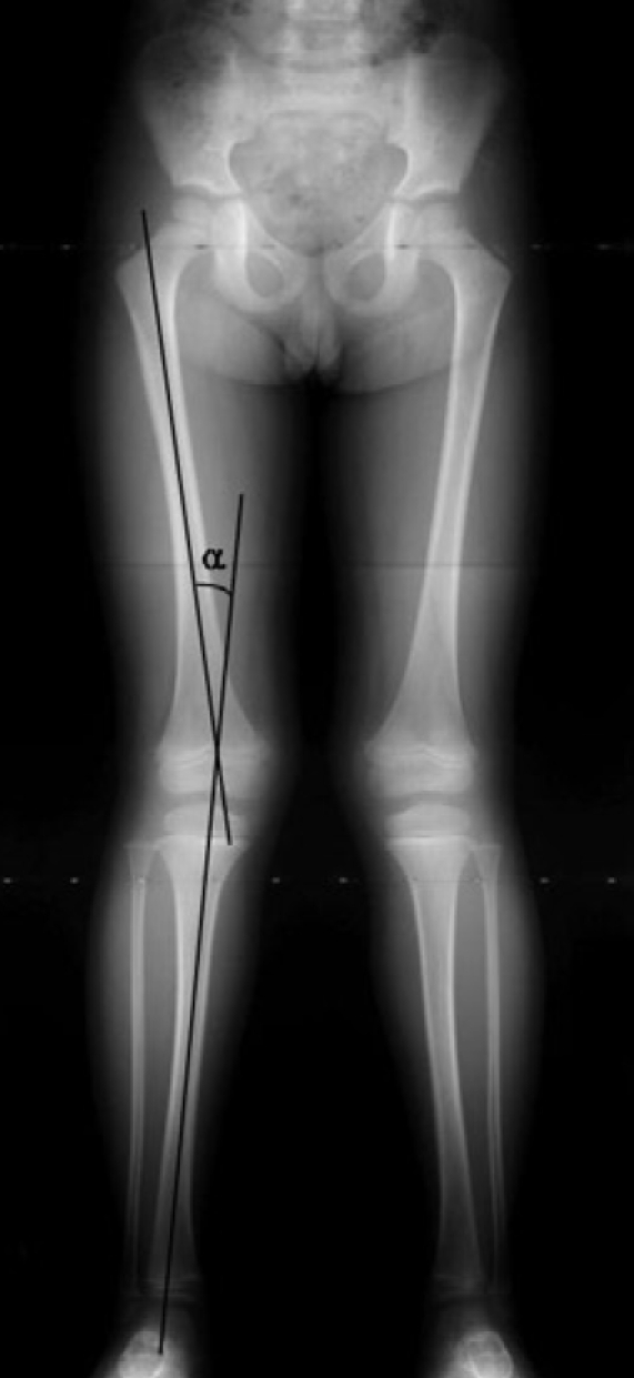
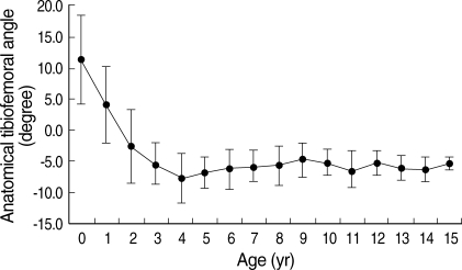
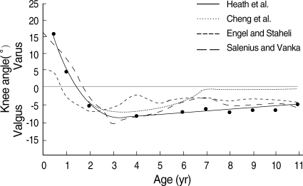
This study was performed to identify the chronological changes of the knee angle or the tibiofemoral angles in normal healthy Korean children. Full-length anteroposterior view standing radiographs of 818 limbs of 452 Korean children were analyzed. The overall patterns of the chronological changes in the knee angle were similar to those described previously in western or Asian children, but the knee angle development was delayed, i.e., genu varum before 1 yr, neutral at 1.5 yr, increasing genu valgum with maximum a value of 7.8 degrees at 4 yr, followed by a gradual decrease to approximately 5-6 degrees of genu valgum of the adult level at 7 to 8 yr of age. These normative data on chronological changes of knee angles should be taken into consideration when evaluating lower limb alignment in children.
Figures
References
-
- McDade W. Bow legs and knock knees. Pediatr Clin North Am. 1977;24:825–839. - PubMed
-
- Sherman M. Physiologic bowing of the legs. South Med J. 1960;53:830–836. - PubMed
-
- Engel GM, Staheli LT. The natural history of torsion and other factors influencing gait in childhood. A study of the angle of gait, tibial torsion, knee angle, hip rotation, and development of the arch in normal children. Clin Orthop Relat Res. 1974;99:12–17. - PubMed
-
- Hachiya M. A roentgenographical study on chronological changes in genu varum and valgum in children (author's transl) Nippon Seikeigeka Gakkai Zasshi. 1981;55:31–43. - PubMed
MeSH terms
LinkOut - more resources
Full Text Sources
