

Benign schwannoma of the liver: a case report
- PMID: 18756066
- PMCID: PMC2526407
- DOI: 10.3346/jkms.2008.23.4.727
Benign schwannoma of the liver: a case report
Abstract
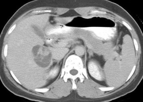
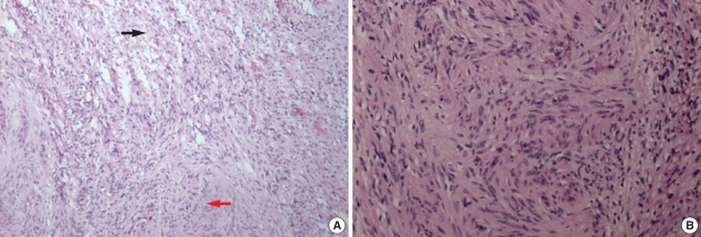
A primary benign schwannoma of the liver is extremely rare. Only nine cases have been reported in the medical literature worldwide and no case has been reported in Korea previously. A 36-yr-old woman was admitted to our hospital with vague epigastric pain. The ultrasound and computed tomography scan revealed a multi-septated cystic mass in the right lobe of the liver. The mass was resected; it was found to be a 5 x 4 x 2 cm mass filled with reddish yellow fluid. The histological examination confirmed the diagnosis of a benign schwannoma, proven by positive immunoreaction with the neurogenic marker S-100 protein and a negative response to CD34, CD117 and smooth muscle actin. This is the first report of a benign schwannoma of the liver parenchyma in a Korean patient.
Figures


References
-
- Pereira Filho RA, Souza SA, Oliveira Filho JA. Primary neurilemmal tumour of the liver: case report. Arq Gastroenterol. 1978;15:136–138. - PubMed
-
- Bekker GM. Neurofibroma of the liver. Sov Med. 1982;10:120–121. - PubMed
-
- Hytiroglou P, Linton P, Klion F, Schwartz M, Miller C, Thung SN. Benign schwannoma of the liver. Arch Pathol Lab Med. 1993;117:216–218. - PubMed
-
- Heffron TG, Coventry S, Bedendo F, Baker A. Resection of primary schwannoma of the liver not associated with neurofibromatosis. Arch Surg. 1993;128:1396–1398. - PubMed
-
- Yoshida M, Nakashima Y, Tanaka A, Mori K, Yamaoka Y. Benign schwannoma of the liver: a case report. Nippon Geka Hokan. 1994;63:208–214. - PubMed
Publication types
MeSH terms
Substances
LinkOut - more resources
Full Text Sources
Medical
