

Narrow-band imaging optical chromocolonoscopy: advantages and limitations
- PMID: 18756593
- PMCID: PMC2739938
- DOI: 10.3748/wjg.14.4867
Narrow-band imaging optical chromocolonoscopy: advantages and limitations
Abstract
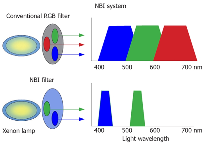
Narrow-band imaging (NBI) is an innovative optical technology that modifies the center wavelength and bandwidth of an endoscope's light into narrow-band illumination of 415 +/- 30 nm. NBI markedly improves capillary pattern contrast and is an in vivo method for visualizing microvessel morphological changes in superficial neoplastic lesions. The scientific basis for NBI is that short wavelength light falls within the hemoglobin absorption band, thereby facilitating clearer visualization of vascular structures. Several studies have reported advantages and limitations of NBI colonoscopy in the colorectum. One difficulty in evaluating results, however, has been non-standardization of NBI systems (Sequential and non-sequential). Utilization of NBI technology has been increasing worldwide, but accurate pit pattern analysis and sufficient skill in magnifying colonoscopy are basic fundamentals required for proficiency in NBI diagnosis of colorectal lesions. Modern optical technology without proper image interpretation wastes resources, confuses untrained endoscopists and delays inter-institutional validation studies. Training in the principles of "optical image-enhanced endoscopy" is needed to close the gap between technological advancements and their clinical usefulness. Currently available evidence indicates that NBI constitutes an effective and reliable alternative to chromocolonoscopy for in vivo visualization of vascular structures, but further study assessing reproducibility and effectiveness in the colorectum is ongoing at various medical centers.
Figures
Similar articles
-
Accuracy of in vivo colorectal polyp discrimination by using dual-focus high-definition narrow-band imaging colonoscopy.Gastrointest Endosc. 2014 Dec;80(6):1072-87. doi: 10.1016/j.gie.2014.05.305. Epub 2014 Jun 25. Gastrointest Endosc. 2014. PMID: 24973171 Clinical Trial.
-
Magnifying endoscopy with narrow band imaging for diagnosis of colorectal tumors.Gastrointest Endosc. 2007 Jun;65(7):988-95. doi: 10.1016/j.gie.2006.07.046. Epub 2007 Feb 26. Gastrointest Endosc. 2007. PMID: 17324407
-
Narrow-band imaging in the colon: limitations and potentials.J Gastroenterol Hepatol. 2011 Nov;26(11):1589-96. doi: 10.1111/j.1440-1746.2011.06877.x. J Gastroenterol Hepatol. 2011. PMID: 21793916 Review.
-
Comparison of white light and narrow band high definition images in predicting colon polyp histology, using standard colonoscopes without optical magnification.Endoscopy. 2008 Oct;40(10):818-22. doi: 10.1055/s-2008-1077437. Epub 2008 Jul 30. Endoscopy. 2008. PMID: 18668472 Clinical Trial.
-
Rationale for and clinical benefits of colonoscopy with narrow band imaging: pathological prediction and colorectal screening.Int J Colorectal Dis. 2013 Jan;28(1):1-7. doi: 10.1007/s00384-012-1591-7. Epub 2012 Oct 9. Int J Colorectal Dis. 2013. PMID: 23053681 Review.
Cited by
-
Assessment of Narrow-Band Imaging Algorithm for Video Capsule Endoscopy Based on Decorrelated Color Space for Esophageal Cancer: Part II, Detection and Classification of Esophageal Cancer.Cancers (Basel). 2024 Jan 29;16(3):572. doi: 10.3390/cancers16030572. Cancers (Basel). 2024. PMID: 38339322 Free PMC article.
-
Clinical usefulness of image-enhanced endoscopy for the diagnosis of ulcerative colitis-associated neoplasia.DEN Open. 2024 Jan 6;4(1):e325. doi: 10.1002/deo2.325. eCollection 2024 Apr. DEN Open. 2024. PMID: 38188357 Free PMC article. Review.
-
How to assess the severity of atrophic gastritis.World J Gastroenterol. 2011 Apr 7;17(13):1690-3. doi: 10.3748/wjg.v17.i13.1690. World J Gastroenterol. 2011. PMID: 21483628 Free PMC article. Review.
-
Trimodal color-fluorescence-polarization endoscopy aided by a tumor selective molecular probe accurately detects flat lesions in colitis-associated cancer.J Biomed Opt. 2014 Dec;19(12):126002. doi: 10.1117/1.JBO.19.12.126002. J Biomed Opt. 2014. PMID: 25473883 Free PMC article.
-
Factors influencing quality of bowel preparation for colonoscopy.World J Gastrointest Endosc. 2013 Feb 16;5(2):39-46. doi: 10.4253/wjge.v5.i2.39. World J Gastrointest Endosc. 2013. PMID: 23424015 Free PMC article.
References
-
- Folkman J. Tumor angiogenesis: therapeutic implications. N Engl J Med. 1971;285:1182–1186. - PubMed
-
- Folkman J, Watson K, Ingber D, Hanahan D. Induction of angiogenesis during the transition from hyperplasia to neoplasia. Nature. 1989;339:58–61. - PubMed
-
- Gono K, Obi T, Yamaguchi M, Ohyama N, Machida H, Sano Y, Yoshida S, Hamamoto Y, Endo T. Appearance of enhanced tissue features in narrow-band endoscopic imaging. J Biomed Opt. 2004;9:568–577. - PubMed
-
- Muto M, Katada C, Sano Y, Yoshida S. Narrow band imaging: a new diagnostic approach to visualize angiogenesis in superficial neoplasia. Clin Gastroenterol Hepatol. 2005;3:S16–S20. - PubMed
-
- Sano Y, Saito Y, Fu KI, Matsuda T, Uraoka T, Kobayashi N, Ito H, Machida H, Iwasaki J, Emura F, et al. Efficacy of Magnifying chromoendoscopy for the differential diagnosis of colorectal lesions. Dig Endosc. 2005;17:105–116.
Publication types
MeSH terms
LinkOut - more resources
Full Text Sources
Other Literature Sources
Medical
Miscellaneous
