. 2008 Oct 31;283(44):30090-100.
doi: 10.1074/jbc.M803290200. Epub 2008 Aug 29.

Gly-Ala repeats induce position- and substrate-specific regulation of 26 S proteasome-dependent partial processing

Affiliations

Gly-Ala repeats induce position- and substrate-specific regulation of 26 S proteasome-dependent partial processing

Chrysoula Daskalogianni et al. J Biol Chem. .

Abstract

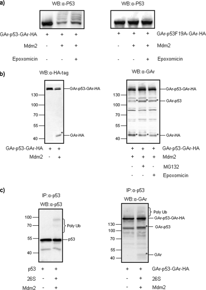
Partial degradation or regulated ubiquitin proteasome-dependent processing by the 26 S proteasome has been demonstrated, but the underlying molecular mechanisms and the prevalence of this phenomenon remain obscure. Here we show that the Gly-Ala repeat (GAr) sequence of EBNA1 affects processing of substrates via the ubiquitin-dependent degradation pathway in a substrate- and position-specific fashion. GAr-mediated increase in stability of proteins targeted for degradation via the 26 S proteasome was associated with a fraction of the substrates being partially processed and the release of the free GAr. The GAr did not cause a problem for the proteolytic activity of the proteasome, and its fusion to the N terminus of p53 resulted in an increase in the rate of degradation of the entire chimera. Interestingly the GAr had little effect on the stability of EBNA1 protein itself, and targeting EBNA1 for 26 S proteasome-dependent degradation led to its complete degradation. Taken together, our data suggest a model in which the GAr prevents degradation or promotes endoproteolytic processing of substrates targeted for the 26 S proteasome by interfering with the initiation step of substrate unfolding. These results will help to further understand the underlying mechanisms for partial proteasome-dependent degradation.


Figures

FIGURE 1.
FIGURE 1.
The GAr does not affect EBNA1 stability. a, H1299 cells were transfected with EBNA1 and the GAr-deleted EBNA1 (EBNAΔGAr) expression constructs. 36 h post-transfection cells were treated with 10 μm cycloheximide (Chx) over an 8-h time course. The figure shows immunoblots using anti-EBNA1 or actin monoclonal antibodies. b, metabolic cell labeling of cells expressing EBNA1 or EBNAΔGAr using 90 μCi of [35S]methionine for 2 h followed by a 12- and 24-h chase in normal medium supplemented with 10 μm methionine. c, H1299 cells were transfected with the Ub-EBNA1 expression construct, and the rate of degradation was analyzed over an 8-h time course in the presence of cycloheximide. Equal loading was confirmed by immunoblotting for actin. All data are representative of at least three independent experiments. WB, Western blot. Error bars represent S.D.
FIGURE 2.
FIGURE 2.
The GAr affects proteasomal degradation in a substrate- and position-dependent way. a, schematic representation of the various GAr-carrying p53 and IκBα constructs. b, the E3 ubiquitin ligase Mdm2 interacts directly with p53 and promotes its polyubiquitination and subsequent degradation by the 26 S proteasome. p53, GAr-p53, and p53-GAr constructs were transfected in H1299 cells in the absence or presence of Mdm2. Transfection efficiency was assayed by co-transfecting cells with green fluorescent protein (GFP), and equal loading was confirmed by immunoblotting for actin. c, an autoradiograph of a 4-h chase experiment following [35S]methionine metabolic pulse labeling of p53 and p53-GAr in the presence of Mdm2. The GAr causes a slowdown of synthesis, and less p53-GAr is therefore labeled (17). Data for the pulse-chase experiment presented in the graph below were obtained using phosphorimager analysis. d, TNFα treatment promotes IκBα degradation by the 26 S proteasome. HA-IκBα wild type (wt), GAr-IκBα, and IκBα-GAr constructs were transfected in HeLa cells. 36 h post-transfection TNFα was added at a final concentration of 50 μm for 30 min to one of each duplicate. Immunoblotting using an α-IκBα monoclonal antibody revealed the levels of endogenous IκBα (IκBα end) and the different exogenous IκBα fusion constructs in the absence (-) or presence (+) of TNFα. Equal loading was confirmed by immunoblotting for actin. The right graph shows the relative amount of protein after TNFα treatment as determined by quantification of Western blot analysis using chemiluminescence and a charge-coupled device camera. Because of differences in expression levels between the constructs before TNFα treatment, the relative amount of each construct is given the arbitrary value of 100, and the relative decrease in expression levels after treatment is shown with S.D. All data are representative of at least three independent experiments. IP, immunoprecipitation; WB, Western blot.
FIGURE 3.
FIGURE 3.
GAr-carrying chimeras are partially processed by the 26 S proteasome. a, H1299 cells expressing p53-GAr were targeted for 26 S-dependent degradation by overexpressing Mdm2. Immunoblotting using an anti-GAr-specific polyclonal serum revealed a band corresponding to the full-length 235-amino acid GAr in cells co-expressing p53-GAr and Mdm2. The F19A-mutated p53 protein cannot be targeted for degradation by Mdm2 (25), and free GAr* was not detected in cells expressing p53F19A-GAr in the presence of Mdm2. The gel was overexposed to show the presence, or absence, of the free GAr product. b, in vitro degradation of p53-GAr using purified 26 S proteasome as described under “Experimental Procedures.” c, fusion of the GAr to the N terminus of p53 (GAr-p53) does not lead to protection from Mdm2-dependent degradation or in the release of the free GAr*. d, HeLa cells expressing the IκBα-GAr fusion protein show an increase in the amount of free GAr* after TNFα treatment that was suppressed in the presence of 20 μm of the proteasome inhibitor MG132. All data are representative of at least three independent experiments. * indicates that the polypeptide is a degradation product. The corresponding less exposed gels are shown in supplemental Fig. 3. wt, wild type; WB, Western blot; IP, immunoprecipitation.
FIGURE 4.
FIGURE 4.
The GAr is a specific regulator of the 26 S proteasome. a, an 80-amino acid-long poly(Q) repeat sequence was fused to p53 (p53-poly(Q) and poly(Q)-p53) and expressed in H1299 cells in the presence or absence of Mdm2. The specific proteasome inhibitor epoxomicin was added 36 h post-transfection at a final concentration of 10 μm for 5 h. b, the release of free poly(Q) cannot be detected using anti-poly(Q) antibodies. Accumulation of polyubiquitinated species is indicated. Equal loading was confirmed by immunoblotting for actin. All data are representative of at least three independent experiments. WB, Western blot.
FIGURE 5.
FIGURE 5.
The 26 S proteasome degrades the N-terminal part of GAr-carrying chimeras. a, fusion of p53 to either end of GAr (HA-p53-GAr-p53) results in degradation of the p53 sequence linked to the N terminus of GAr and the release of the free GAr-p53*. Treatment with epoxomicin for 5 h results in increased levels of HA-p53-GAr-p53, whereas GAr-p53* levels are slightly decreased. The graph shows the quantification of the corresponding immunoblot (upper left panel). The levels of the triple chimera do not change when cells are treated with several other protease inhibitors (middle panel). The HA-GAr-p53 polypeptide expressed from the HA-GAr-p53 mRNA was detected using an anti-HA tag antibody, but the GAr-p53* that is derived from degradation of the HA-p53-GAr-p53 was not (right panel). b, an autoradiograph showing a 30-min [35S]methionine pulse label of cells expressing HA-GAr-p53 and HA-p53-GAr-p53 in the presence or absence of MG132 followed by an immunoprecipitation (IP) using an anti-HA tag monoclonal antibody. c, HA-IκBα-GAr-p53 fusion protein is partially degraded by the 26 S proteasome. Both the triple HA-IκBα-GAr-p53 and the GAr-p53* product are targeted for degradation by Mdm2 (left panel). When cells are treated with TNFα only the triple construct is targeted for degradation, leading to an accumulation of GAr-p53* (right panel). Treatment with epoxomicin prevents Mdm2- or TNFα-dependent degradation of HA-IκBα-GAr-p53 and results in less GAr-p53*. The graphs show the quantification of the corresponding Western blots (WB), and the HA-IκBα-GAr-p53 in the absence of Mdm2 or TNFα has been given the arbitrary value 100. All data are representative of at least three independent experiments. * indicates that the polypeptide is a degradation product. AEBSF, 4-(2-aminoethyl)benzenesulfonyl fluoride. Error bars represent S.D.
FIGURE 6.
FIGURE 6.
Partial processing of GAr-carrying chimeras by the 26 S proteasome is preceded by an endoproteolytic cleavage. GAr was fused to either end of p53 (GAr-p53-GAr-HA) to protect the substrate from end first-mediated degradation by the proteasome. a, GAr-p53-GAr-HA or GAr-p53F19A-GAr-HA expression constructs were transfected in H1299 cells in the absence or presence of Mdm2 and treated with epoxomicin where indicated. b, co-expression of Mdm2 targets the triple chimera for degradation and results in the appearance of GAr-p53* and GAr-HA* degradation products (left and right panels). Immunoblotting using an anti-HA tag antibody confirms the identity of the degradation products (left panel). Treatment with the proteasome inhibitors MG132 or epoxomicin leads to a decrease in the amount of GAr-HA* (right panel). c, in vitro degradation of p53 or GAr-p53-GAr-HA using immunopurified substrates, 26 S proteasome, and Mdm2 as described under “Experimental Procedures.” All data are representative of at least three independent experiments. * indicates that the polypeptide is a degradation product. WB, Western blot; IP, immunoprecipitation.
FIGURE 7.
FIGURE 7.
Model for GAr-dependent interference with the 26 S proteasome. Fusion of GAr far from the natural unfolding tag of a protein (e.g. in the N terminus of p53) does not prevent 26 S-dependent degradation and results in the complete degradation of the entire chimera by the 26 S proteasome. When GAr is instead fused next to the unfolding tag it impairs unfolding of the substrate, leading to an inhibition of degradation of the majority of the chimeras. In a small fraction of GAr chimeras, however, a loop structure is formed at the unfolding tag and is threaded into the 20 S chamber and endoproteolytically cleaved, leading to the release of GAr fragments and degradation of the rest of the substrate. This model predicts that the GAr is affecting the unfolding of the substrate that takes place at the 19 S regulatory subunit before processing by the 20 S core particle can start. Hence it can explain how the GAr (i) has no effect or (ii) prevents degradation and causes partial processing of the same substrate depending on its location.

Similar articles

Cited by

References

    1. Glickman, M. H., and Ciechanover, A. (2002) Physiol. Rev. 82 373-428 - PubMed
    1. Elsasser, S., and Finley, D. (2005) Nat. Cell Biol. 7 742-749 - PubMed
    1. Rape, M., and Jentsch, S. (2002) Nat. Cell Biol. 4 E113-E116 - PubMed
    1. Hoppe, T., Matuschewski, K., Rape, M., Schlenker, S., Ulrich, H. D., and Jentsch, S. (2000) Cell 102 577-586 - PubMed
    1. Hoppe, T., Rape, M., and Jentsch, S. (2001) Curr. Opin. Cell Biol. 13 344-348 - PubMed

Publication types