

In vivo analysis of protein kinase B (PKB)/Akt regulation in DNA-PKcs-null mice reveals a role for PKB/Akt in DNA damage response and tumorigenesis
- PMID: 18757368
- PMCID: PMC2662067
- DOI: 10.1074/jbc.M803053200
In vivo analysis of protein kinase B (PKB)/Akt regulation in DNA-PKcs-null mice reveals a role for PKB/Akt in DNA damage response and tumorigenesis
Abstract
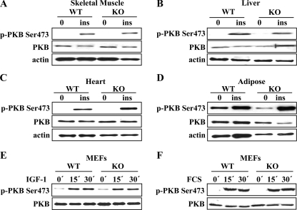
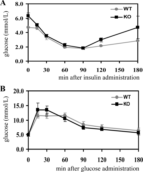
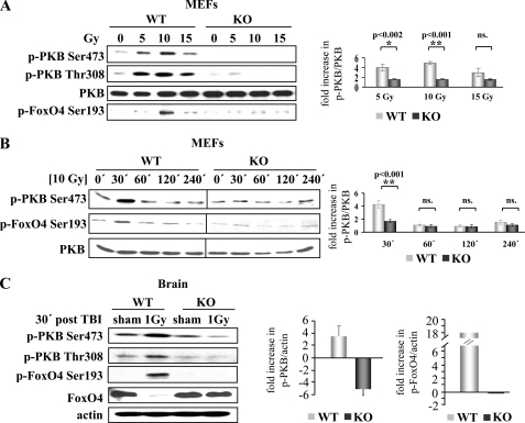
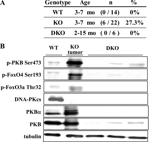
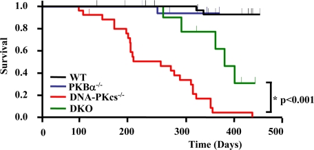
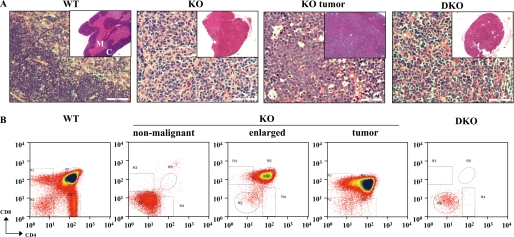
Full activation of protein kinase B (PKB/Akt) requires phosphorylation on Thr-308 and Ser-473. It is well established that Thr-308 is phosphorylated by 3-phosphoinositide-dependent kinase-1 (PDK1). Ser-473 phosphorylation is mediated by both mammalian target of rapamycin-rictor complex (mTORC2) and DNA-dependent protein kinase (DNA-PK) depending on type of stimulus. However, the physiological role of DNA-PK in the regulation of PKB phosphorylation remains to be established. To address this, we analyzed basal, insulin-induced, and DNA damage-induced PKB Ser-473 phosphorylation in DNA-PK catalytic subunit-null DNA-PKcs(-/-) mice. Our results revealed that DNA-PK is required for DNA damage-induced phosphorylation but dispensable for insulin- and growth factor-induced PKB Ser-473 phosphorylation. Moreover, DNA-PKcs(-/-) mice showed a tissue-specific increase in basal PKB phosphorylation. In particular, persistent PKB hyperactivity in the thymus apparently contributed to spontaneous lymphomagenesis in DNA-PKcs(-/-) mice. Significantly, these tumors could be prevented by deletion of PKBalpha. These findings reveal stimulus-specific regulation of PKB activation by specific upstream kinases and provide genetic evidence of PKB deregulation in DNA-PKcs(-/-) mice.
Figures

Similar articles
-
DNA-dependent protein kinase catalytic subunit (DNA-PKcs)-SIN1 association mediates ultraviolet B (UVB)-induced Akt Ser-473 phosphorylation and skin cell survival.Mol Cancer. 2013 Dec 24;12(1):172. doi: 10.1186/1476-4598-12-172. Mol Cancer. 2013. PMID: 24365180 Free PMC article.
-
Identification of a PKB/Akt hydrophobic motif Ser-473 kinase as DNA-dependent protein kinase.J Biol Chem. 2004 Sep 24;279(39):41189-96. doi: 10.1074/jbc.M406731200. Epub 2004 Jul 15. J Biol Chem. 2004. PMID: 15262962
-
PKBalpha/Akt1 acts downstream of DNA-PK in the DNA double-strand break response and promotes survival.Mol Cell. 2008 Apr 25;30(2):203-13. doi: 10.1016/j.molcel.2008.02.024. Mol Cell. 2008. PMID: 18439899
-
PIKKing on PKB: regulation of PKB activity by phosphorylation.Curr Opin Cell Biol. 2009 Apr;21(2):256-61. doi: 10.1016/j.ceb.2009.02.002. Epub 2009 Mar 19. Curr Opin Cell Biol. 2009. PMID: 19303758 Review.
-
Protein kinase B (PKB/Akt), a key mediator of the PI3K signaling pathway.Curr Top Microbiol Immunol. 2010;346:31-56. doi: 10.1007/82_2010_58. Curr Top Microbiol Immunol. 2010. PMID: 20517722 Review.
Cited by
-
Akt kinase C-terminal modifications control activation loop dephosphorylation and enhance insulin response.Biochem J. 2015 Oct 1;471(1):37-51. doi: 10.1042/BJ20150325. Epub 2015 Jul 22. Biochem J. 2015. PMID: 26201515 Free PMC article.
-
Repair of radiation damage of U2OS osteosarcoma cells is related to DNA-dependent protein kinase catalytic subunit (DNA-PKcs) activity.Mol Cell Biochem. 2014 May;390(1-2):51-9. doi: 10.1007/s11010-013-1955-5. Epub 2014 Jan 5. Mol Cell Biochem. 2014. PMID: 24390088
-
Transcriptome analysis of the circadian clock gene BMAL1 deletion with opposite carcinogenic effects.Funct Integr Genomics. 2021 Jan;21(1):1-16. doi: 10.1007/s10142-020-00757-6. Epub 2020 Oct 27. Funct Integr Genomics. 2021. PMID: 33111200
-
DNA-PK mediates AKT activation and apoptosis inhibition in clinically acquired platinum resistance.Neoplasia. 2011 Nov;13(11):1069-80. doi: 10.1593/neo.111032. Neoplasia. 2011. PMID: 22131882 Free PMC article.
-
Role of AKT signaling in DNA repair and clinical response to cancer therapy.Neuro Oncol. 2014 Oct;16(10):1313-23. doi: 10.1093/neuonc/nou058. Epub 2014 May 7. Neuro Oncol. 2014. PMID: 24811392 Free PMC article. Review.
References
-
- Brazil, D. P., and Hemmings, B. A. (2001) Trends Biochem. Sci. 26 657-664 - PubMed
-
- Fayard, E., Tintignac, L. A., Baudry, A., and Hemmings, B. A. (2005) J. Cell Sci. 118 5675-5678 - PubMed
-
- Parcellier, A., Tintignac, L. A., Zhuravleva, E., and Hemmings, B. A. (2008) Cell. Signal. 20 21-30 - PubMed
-
- Greer, E. L., and Brunet, A. (2005) Oncogene 24 7410-7425 - PubMed
Publication types
MeSH terms
Substances
LinkOut - more resources
Full Text Sources
Molecular Biology Databases
Miscellaneous
