

Tetrasulfated disaccharide unit in heparan sulfate: enzymatic formation and tissue distribution
- PMID: 18757372
- PMCID: PMC2662186
- DOI: 10.1074/jbc.M801586200
Tetrasulfated disaccharide unit in heparan sulfate: enzymatic formation and tissue distribution
Abstract
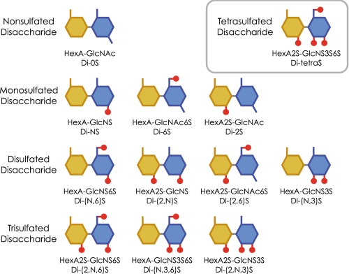
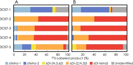
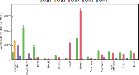
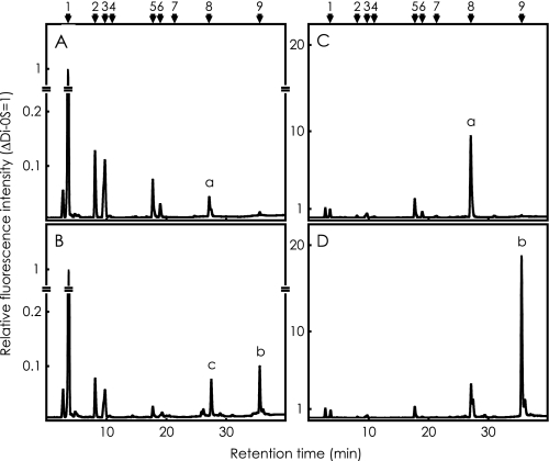
We previously reported that the heparan sulfate 3-O-sulfotransferase (3OST)-5 produces a novel component of heparan sulfate, i.e. the tetrasulfated disaccharide (Di-tetraS) unit ( Mochizuki, H., Yoshida, K., Gotoh, M., Sugioka, S., Kikuchi, N., Kwon, Y.-D., Tawada, A., Maeyama, K., Inaba, N., Hiruma, T., Kimata, K., and Narimatsu, H. (2003) J. Biol. Chem. 278, 26780-26787 ). In the present study, we investigated the potential of other 3OST isoforms to produce Di-tetraS with heparan sulfate and heparin as acceptor substrates. 3OST-2, 3OST-3, and 3OST-4 produce Di-tetraS units as a major product from both substrates. 3OST-5 showed the same specificity for heparin, but the production from heparan sulfate was very low. Di-tetraS production by 3OST-1 was negligible. We then investigated the presence of Di-tetraS units in heparan sulfates from various rat tissues. Di-tetraS was detected in all of the tissues analyzed. Liver and spleen contain relatively high levels of Di-tetraS, 1.6 and 0.95%, respectively. However, the content of this unit in heart, large intestine, ileum, and lung is low, less than 0.2%. We further determined the expression levels of 3OST transcripts by quantitative real time PCR. The 3OST-3 transcripts are highly expressed in spleen and liver. The 3OST-2 and -4 are specifically expressed in brain. These results indicate that the Di-tetraS unit is widely distributed throughout the body as a rare and unique component of heparan sulfate and is synthesized by tissue-specific 3OST isoforms specific for Di-tetraS production.
Figures
Similar articles
-
Characterization of a heparan sulfate 3-O-sulfotransferase-5, an enzyme synthesizing a tetrasulfated disaccharide.J Biol Chem. 2003 Jul 18;278(29):26780-7. doi: 10.1074/jbc.M301861200. Epub 2003 May 9. J Biol Chem. 2003. PMID: 12740361
-
A quantitative method to detect non-antithrombin-binding 3-O-sulfated units in heparan sulfate.J Biol Chem. 2021 Jan-Jun;296:100115. doi: 10.1074/jbc.RA120.015864. Epub 2020 Dec 3. J Biol Chem. 2021. PMID: 33234593 Free PMC article.
-
NDST2 (N-Deacetylase/N-Sulfotransferase-2) Enzyme Regulates Heparan Sulfate Chain Length.J Biol Chem. 2016 Sep 2;291(36):18600-18607. doi: 10.1074/jbc.M116.744433. Epub 2016 Jul 7. J Biol Chem. 2016. PMID: 27387504 Free PMC article.
-
Use of biosynthetic enzymes in heparin and heparan sulfate synthesis.Bioorg Med Chem. 2013 Aug 15;21(16):4786-92. doi: 10.1016/j.bmc.2012.11.053. Epub 2012 Dec 12. Bioorg Med Chem. 2013. PMID: 23313092 Review.
-
Heparan sulfate and development: differential roles of the N-acetylglucosamine N-deacetylase/N-sulfotransferase isozymes.Biochim Biophys Acta. 2002 Dec 19;1573(3):209-15. doi: 10.1016/s0304-4165(02)00386-0. Biochim Biophys Acta. 2002. PMID: 12417402 Review.
Cited by
-
Sex Difference Leads to Differential Gene Expression Patterns and Therapeutic Efficacy in Mucopolysaccharidosis IVA Murine Model Receiving AAV8 Gene Therapy.Int J Mol Sci. 2022 Oct 21;23(20):12693. doi: 10.3390/ijms232012693. Int J Mol Sci. 2022. PMID: 36293546 Free PMC article.
-
Synthesis of heparan sulfate with cyclophilin B-binding properties is determined by cell type-specific expression of sulfotransferases.J Biol Chem. 2010 Jan 15;285(3):1701-15. doi: 10.1074/jbc.M109.018184. Epub 2009 Nov 23. J Biol Chem. 2010. PMID: 19940140 Free PMC article.
-
Heparinase Digestion of 3-O-Sulfated Sequences: Selective Heparinase II Digestion for Separation and Identification of Binding Sequences Present in ATIII Affinity Fractions of Bovine Intestinal Heparins.Front Med (Lausanne). 2022 Mar 31;9:841726. doi: 10.3389/fmed.2022.841726. eCollection 2022. Front Med (Lausanne). 2022. PMID: 35433769 Free PMC article.
-
Development of Substrate Degradation Enzyme Therapy for Mucopolysaccharidosis IVA Murine Model.Int J Mol Sci. 2019 Aug 24;20(17):4139. doi: 10.3390/ijms20174139. Int J Mol Sci. 2019. PMID: 31450640 Free PMC article.
-
3-O-Sulfation induces sequence-specific compact topologies in heparan sulfate that encode a dynamic sulfation code.Comput Struct Biotechnol J. 2022 Jul 18;20:3884-3898. doi: 10.1016/j.csbj.2022.07.013. eCollection 2022. Comput Struct Biotechnol J. 2022. PMID: 35891779 Free PMC article.
References
-
- Yanagishita, M., and Hascall, V. C. (1992) J. Biol. Chem. 2679451 -9454 - PubMed
-
- Bernfield, M., Götte, M., Park, P. W., Reizes, O., Fitzgerald, M. L., Lincecum, J., and Zako, M. (1999) Annu. Rev. Biochem. 68729 -777 - PubMed
-
- Esko, J. D., and Selleck, S. B. (2002) Annu. Rev. Biochem. 71435 -471 - PubMed
-
- Lin, X. (2004) Development 1316009 -6021 - PubMed
-
- Bishop, J. R., Schuksz, M., and Esko, J. D. (2007) Nature 4461030 -1037 - PubMed
MeSH terms
Substances
LinkOut - more resources
Full Text Sources
Other Literature Sources
Molecular Biology Databases
