. 2008 Sep 1;68(17):6963-8.
doi: 10.1158/0008-5472.CAN-08-0627.

EBV is necessary for proliferation of dually infected primary effusion lymphoma cells

Affiliations

EBV is necessary for proliferation of dually infected primary effusion lymphoma cells

Amanda A Mack et al. Cancer Res. .

Abstract

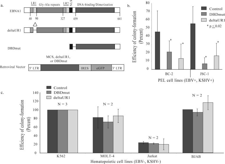
Epstein Barr virus (EBV) and Kaposi's sarcoma-associated herpesvirus (KSHV) are found together in approximately 80% of primary effusion lymphomas (PEL), but their contribution to these cancers is unclear. We found that dominant-negative derivatives of EBNA1 inhibited EBV-positive PEL cells from forming colonies. Those rare PEL cells that proliferated after expression of the dominant-negative derivatives usually expressed these derivatives at low or undetectable levels and continued to maintain their EBV genomes. Those proliferating cells expressing higher levels of the derivatives expressed mutant derivatives that could not bind DNA. These findings indicate that EBV is required to sustain proliferation, as measured by colony formation of dually infected PEL cells. The dominant-negative derivatives of EBNA1 had no effect on the colony-forming ability of five EBV-negative, KSHV-negative hematopoietic cell lines. Surprisingly, they did inhibit the colony-forming ability of EBV-negative, KSHV-positive PEL cells. The small fraction of cells that continued to proliferate expressed only mutants of the EBNA1 derivatives that could no longer bind DNA. These findings indicate that the site-specific DNA-binding activity of EBNA1 or its derivatives when expressed efficiently in EBV-negative, KSHV-positive PEL cells inhibits their colony formation possibly through their binding to the KSHV genome.


Figures

Figure 1
Figure 1. Two derivatives of EBNA1 inhibit EBV+, KSHV+ PEL cells from forming colonies and do not affect EBV−, KSHV− hematopoietic cell lines
(a) Full-length EBNA1 is 641 amino acids in length, contains two regions (LR1 and LR2) responsible for linking DNA, a nuclear localization sequence (NLS) and a DNA-binding and dimerization domain (15). This carboxy-terminal DNA-binding and dimerization domain is not sufficient to support EBNA1's contributions to DNA replication and acts as a dominant-negative derivative inhibiting all of these functions (9, 15). Numbers indicate the position of amino acids. The deletion within deltaUR1 spans amino acids 65 − 89 (Delta) rendering EBNA1 transcriptionally defective. DBDmut contains only an NLS and the DNA-binding and dimerization domain and is defective in all of EBNA1's known functions. DBDmut, deltaUR1, or a multiple cloning sequence (MCS) as the control was inserted into a retroviral backbone as previously described (9) that co-expresses eGFP. (b) BC-2 and JSC-1 cells were infected with retroviruses expressing DBDmut, deltaUR1, or the control retrovirus containing only the MCS. Infected cells were sorted by FACs 48 hours post-infection, and their efficiency of forming colonies determined 2 weeks post-plating. (c) DBDmut and deltaUR1 do not inhibit EBV(−), KSHV(−) hematopoietic cell lines from forming colonies. Two EBV-negative Burkitt's lymphoma cell lines BJAB and DG75 (analyzed in reference 9); an erythroleukemia cell line (K562); and two T-cell lymphoblastic leukemia lines (MOLT-4 and Jurkat) were tested in assays for colony-formation. These cells were infected with control, DBDmut, or deltaUR1-expressing retroviruses, sorted into 96-well plates and measured for their ability to form colonies 2 weeks post-plating. N, number of independently performed experiments.
Figure 2
Figure 2. Only JSC-1 cells expressing mutant forms of deltaUR1 and retaining EBV survive to form colonies
(a) Shown is an illustration depicting the two different cell populations analyzed by Western blotting. The Parental population is the initial set of cells transduced with the highest levels of eGFP-expression while the Survivors are the transduced cells that successfully formed the clones recovered from colony-formation assays. (b) Six lysates chosen from a total of fourteen deltaUR1-JSC-1 clones that were the Survivors were analyzed by Western blotting and compared with the corresponding Parental populations, B and C. 3 × 10⁴ cells were loaded in each lane. 721 cells were loaded as an EBNA1-positive control and tubulin served as a loading control. (c) Clones of Survivors have distributions of the number of EBV genomes per cell similar to that of uninfected JSC-1 cells. The number of viral DNAs per cell was measured by FISH. These distributions for three representative clones of Survivors are shown (B.1, B.3, and C.1) along with that of uninfected JSC-1 cells.
Figure 3
Figure 3. Levels of EBNA1 are atypically low in three EBV-positive PEL cell lines
Lysates from three EBV-infected PEL cell lines JSC-1, BC-2, and BC-1 were examined by Western blotting to determine the levels of EBNA1. The intensities of the signals for EBNA1 were determined using ImageQuant software version 5.2. The numbers at the bottom of each lane in the Western blots represent the amount of EBNA1 relative to the levels found in EBV-positive 721 cells. Antibody specificity was determined using the EBV-negative cell lines BJAB and BC-3 as well as the EBV-positive cell line 721. The size of EBNA1 detected in each of these cell lines fell within the estimated 72 kD range as indicated by the size marker located in the first lane of each panel.
Figure 4
Figure 4. EBV(−), KSHV(+) PEL cell lines are inhibited in their ability to form colonies by derivatives of EBNA1 and only BC-3 cells with mutant forms of these derivatives survive to form colonies
(a) BCBL-1, BCP-1, and BC-3 cells were infected with retroviruses expressing DBDmut, deltaUR1, or the control retrovirus. Infected cells were sorted by FACs for the highest levels of eGFP, 5 − 10% of the total population, 48 hours post-infection into 96-well plates. Their colony-forming efficiency was determined using a Poisson Distribution. (b) KSHV DNA was detected in EBV−, KSHV+ BC-3 cells by FISH and no difference in the number of KSHV genomes in the presence or absence of deltaUR1 expression 10 days post-infection was observed. (c) Six lysates of Survivors from deltaUR1-BC-3 clones were analyzed by Western blotting and compared with a corresponding Parental population. Lysates from 3 × 10⁴ cells were loaded for each lane representing Survivors and 1 × 10⁴ cells for the Parent population. 1 × 10⁴ (1x) and 3 × 10⁴ (3x) 721 cells were loaded as an EBNA1-positive control and tubulin was included as a loading control.

References

    1. Jenner RG, Boshoff C. The molecular pathology of Kaposi's sarcoma-associated herpesvirus. Biochim Biophys Acta. 2002;1602:1–22. - PubMed
    1. Walts AE, Shintaku IP, Said JW. Diagnosis of malignant lymphoma in effusions from patients with AIDS by gene rearrangement. Am J Clin Pathol. 1990;94:170–5. - PubMed
    1. Knowles DM, Inghirami G, Ubriaco A, Dalla-Favera R. Molecular genetic analysis of three AIDS-associated neoplasms of uncertain lineage demonstrates their B-cell derivation and the possible pathogenetic role of the Epstein-Barr virus. Blood. 1989;73:792–9. - PubMed
    1. Green I, Espiritu E, Ladanyi M, et al. Primary lymphomatous effusions in AIDS: a morphological, immunophenotypic, and molecular study. Mod Pathol. 1995;8:39–45. - PubMed
    1. Cesarman E, Chang Y, Moore PS, Said JW, Knowles DM. Kaposi's sarcoma-associated herpesvirus-like DNA sequences in AIDS-related body-cavity-based lymphomas. N Engl J Med. 1995;332:1186–91. - PubMed

Publication types

MeSH terms

Substances