. 2008 Sep 1;68(17):7100-9.
doi: 10.1158/0008-5472.CAN-07-6145.

EWS-FLI1 suppresses NOTCH-activated p53 in Ewing's sarcoma

Affiliations

EWS-FLI1 suppresses NOTCH-activated p53 in Ewing's sarcoma

Jozef Ban et al. Cancer Res. .

Abstract

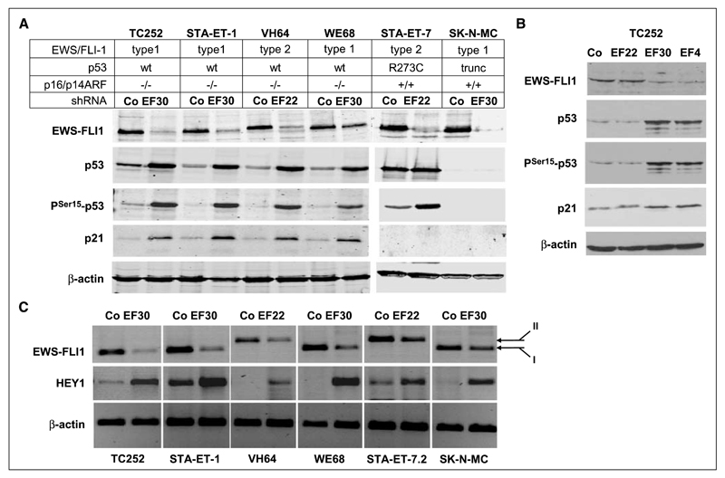
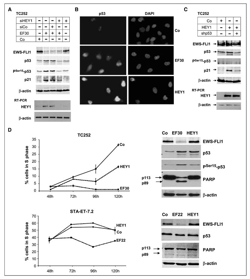
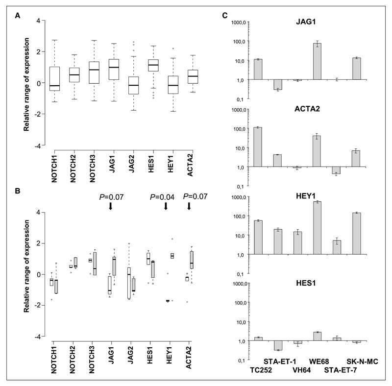
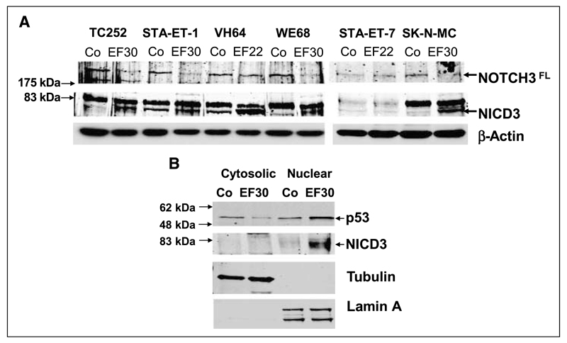
Although p53 is the most frequently mutated gene in cancer, half of human tumors retain wild-type p53, whereby it is unknown whether normal p53 function is compromised by other cancer-associated alterations. One example is Ewing's sarcoma family tumors (ESFT), where 90% express wild-type p53. ESFT are characterized by EWS-FLI1 oncogene fusions. Studying 6 ESFT cell lines, silencing of EWS-FLI1 in a wild-type p53 context resulted in increased p53 and p21(WAF1/CIP1) levels, causing cell cycle arrest. Using a candidate gene approach, HEY1 was linked to p53 induction. HEY1 was rarely expressed in 59 primary tumors, but consistently induced upon EWS-FLI1 knockdown in ESFT cell lines. The NOTCH signaling pathway targets HEY1, and we show NOTCH2 and NOTCH3 to be expressed in ESFT primary tumors and cell lines. Upon EWS-FLI1 silencing, NOTCH3 processing accompanied by nuclear translocation of the activated intracellular domain was observed in all but one p53-mutant cell line. In cell lines with the highest HEY1 induction, NOTCH3 activation was the consequence of JAG1 transcriptional induction. JAG1 modulation by specific siRNA, NOTCH-processing inhibition by either GSI or ectopic NUMB1, and siRNA-mediated HEY1 knockdown all inhibited p53 and p21(WAF1/CIP1) induction. Conversely, forced expression of JAG1, activated NOTCH3, or HEY1 induced p53 and p21(WAF1/CIP1). These results indicate that suppression of EWS-FLI1 reactivates NOTCH signaling in ESFT cells, resulting in p53-dependent cell cycle arrest. Our data link EWS-FLI1 to the NOTCH and p53 pathways and provide a plausible basis both for NOTCH tumor suppressor effects and oncogenesis of cancers that retain wild-type p53.


Conflict of interest statement

Disclosure of Potential Conflicts of Interest

No potential conflicts of interest were disclosed.

Figures

Figure 1
Figure 1
EWS-FLI1 silencing induces expression of activated p53, p21WAF1/CIP1, and HEY1 expression in wild-type p53 ESFT cell lines. A, p53, PSer15-p53, and p21WAF1/CIP1 accumulation after EWS-FLI1 silencing in four wt-p53 and two mt-p53 ESFT cell lines. Immunoblot analysis 4 d after transfection of the indicated shRNA expression vectors, demonstrating induction of Ser 15-phosphorylated p53 and p21WAF1/CIP1 restricted to the wt-p53 cell lines upon introduction of shRNA specific for the respective EWS-FLI1 fusion types (EF30 for type 1 EWS-FLI1 in TC252, STA-ET-1, WE68, and SK-N-MC cells; EF22 for type 2 EWS-FLI1 in VH64 and STA-ET-7.2 cells) compared with control transfections with a nontargeting shRNA (Co). β-Actin is shown as a loading control. B, p53, PSer15-p53, and p21WAF1/CIP1 accumulation are specifically associated with EWS-FLI1–modulating shRNAs. TC252 cells were transfected with either nontargeting shRNA, shRNAs targeting the EWS-FLI1 type 1 fusion region (EF30) or the FLI1 3′ portion (EF4), or with a shRNA to the type 2 fusion region as a mismatched control. EWS-FLI1 and p53 protein expression was monitored by Western blotting. C, demonstration of HEY1 induction upon EWS-FLI1 silencing by RT-PCR 96 h after EWS-FLI1 silencing. For control, a nontargeting shRNA was transfected. β-actin PCR was used as a control for equal input. EWS-FLI1 fusion types I and II are indicated.
Figure 2
Figure 2
Induction of p53 and p21WAF1/CIP1 is dependent on HEY1 expression and associated with cell cycle arrest. A, TC252 cells were transfected with either a nontargeting shRNA or EF30, in the absence or presence of a control nontargeting siRNA pool (siCo) or a siRNA to HEY1 (siHEY1), as indicated. Protein expression of EWS-FLI1, p53, PSer15-p53, p21, and β-actin for loading control was monitored on the Western blot; HEY1 expression was followed on the RNA level by RT-PCR. B, nuclear accumulation of p53 in TC252 cells upon EWS-FLI1 silencing (EF30) and ectopic HEY1 expression (HEY1), monitored by immunostaining with the p53-specific mAb DO1. For control, cells were tansfected with a nontargeting shRNA. Left, p53 staining; right, 4′,6-diamidino-2-phenylindole staining of nuclei. C, HEY1-mediated induction of p21WAF1/CIP1 expression is dependent on the presence of p53. TC252 cells were transfected with either 4-μg pSPORT empty vector (Co) or pSPORT-based HEY1 expression vector in the absence or presence of 1.5 μg of shp53. p53, PSer15-p53, p21, and β-actin protein expression were followed on the Western blot 96 h after transfection. Expression of the HEY1 transgene was monitored by RT-PCR. Note that in contrast to EWS-FLI1 silencing (Fig. 1), HEY1 does not induce p53 phosphorylation. D, cell fate determination after EWS-FLI1 silencing or ectopic HEY1 expression compared with control transfection. TC252 cells and STA-ET-7.2 cells were transfected with the indicated EWS-FLI1 silencing vectors (EF30 or EF22) or with a nontargeting shRNA in duplicates, subjected to puromycin selection 24 h after transfection, and the fraction of surviving cells incorporating the nucleoside analogue EdU within a 45-min incubation period was monitored in 24 h intervals over 4 d starting 48 h after transfection. EdU incorporation was measured by flow cytometry, exclusively gating on living cells. At 96 h posttransfection, protein extracts were prepared from all puromycin-resistant cells and tested for EWS-FLI1, p53, and PARP expression. The reduction of the p113 full length PARP band and the occurrence of a p89 PARP cleavage band were taken as an indication for apoptosis.
Figure 3
Figure 3
Range of relative expression values for selected NOTCH pathway genes shown as boxplots of (A) 3 pooled primary ESFT data sets (18–20; 59 tumors) and (B) 5 ESFT cell lines before (control-treated for 96 h, white bars) and after silencing of EWS-FLI1 (EF-treated for 96 h, gray bars). Expression values are standardized relative to the mean of 79 tissues in the Novartis Gene Expression Atlas, so that zero denotes the mean expression across all analyzed tissues. Therefore, for a given gene in the plot, a deviation from zero indicates that this gene is expressed at higher or lower levels in ESFT cell lines or primary tumors than in the tissue collection, and this deviation can be compared across different genes. Boxes, middle 50% of the values; black horizontal bars, median; the ‘‘whiskers’’ extend to the most extreme values not defined as outliers (1.5 interquartile range from the box); gray circles, outliers. In the cell lines, changes in gene expression upon EWS-FLI1 silencing were significant for HEY1 and marginally significant for JAG1 and ACTA2 with the indicated P values. C, qRT-PCR results for NOTCH pathway genes JAG1, ACTA2, HEY1, and HES1 in the six ESFT cell lines comparing EWS-FLI1 knockdown with control-treated (nontargeting shRNA) cells. Results are presented as fold change on a logarithmic scale; columns, mean relative values of triplicate experiments normalized for β2 microglobulin expression; bars, SD.
Figure 4
Figure 4
Silencing of EWS-FLI1 induces NOTCH3 activation. A, protein extracts from the EWS-FLI1 knockdown experiment presented in Fig. 1 were probed for the expression of the 250-kDa full-length NOTCH3 (NOTCH3FL) and the presence of the ~80-kDa processed intracellular domain (NICD3). B, EWS-FLI1 silencing induces translocation of processed NICD3 to the nucleus. Control nontargeting shRNA and EF30-transfected TC252 were subjected to subcellular fractionation 96 h after transfection. Cytosolic and nuclear extracts were probed for the presence of processed NICD3 and p53. The purity of the extracts was tested using antibodies to cytoplasmic α-tubulin and nuclear lamin A.
Figure 5
Figure 5
EWS-FLI1–induced NOTCH pathway activation induces p53 and p21WAF1/CIP1 in TC252 cells. A, immunoblot monitoring NOTCH3 processing, PSer15-p53 and p21 induction in the absence and presence of 10 μmol/L GSI in two ESFT cell lines (TC252 and WE68) transfected with either nontargeting shRNA or EF30. Note that GSI reduced overall p53 expression, whereas Ser15 phosphorylation was not affected. B, cotransfection of siRNA to JAG1 or of a NUMB expression vector with EF30 reverts p53 and p21WAF1/CIP1 abundancy to control levels, whereas ectopic JAG1 and DLL1 induce p53 and p21 similar to EWS-FLI1 silencing. For negative control, transfection of a nontargeting shRNA (left) and of an empty expression vector (right) are shown. NICD3 transfection served as a positive control. C, regulation of JAG1 in response to EWS-FLI1 silencing is confined to a 570-bp genomic region upstream of the JAG1 transcription initiation site. Firefly luciferase reporter gene assays for a 0.57-kb JAG1 promoter fragment were performed in three ESFT cell lines (TC252, SK-N-MC, and WE68) transfected with EF30 or control (empty vector or nontargeting shRNA). Fold changes in the transcriptional activity of the JAG1 promoter sequences after EWS-FLI1 knockdown were calculated as follows: Foldchange=[574/1JAG1.Luc/TKp.rhL]EWSFLI1shRNA/[574/1JAG1.Luc/TKp.rhL]ScrambledshRNA[TKp.Luc/TKp.rhL]EWS-FLI1shRNA/[TKp.Luc/TKp.rhL]ScrambledshRNA Mean values for fold change from three to five independent experiments, each performed in triplicate are shown. D, ectopic expression of JAG1 in the presence of either siRNA to HEY1 or of GSI reverts p53 abundancy almost to control levels.
Figure 6
Figure 6
Model for p53 and p21WAF1/CIP1 regulation by EWS-FLI1 in TC252 cells. Suppression of EWS-FLI1 results in JAG1 expression by a thus far unknown mechanism. Presumably intercellular interaction between JAG1 and constitutively expressed NOTCH3 (and possibly also NOTCH2) results in γ-secretase–mediated NOTCH cleavage. The NICD translocates to the nucleus and activates CBF/CSL-dependent HEY1 expression. This leads to posttranscriptional stabilization of p53 via a thus far undefined mechanism, and p53-dependent p21WAF1/CIP1 expression. There is evidence that HEY1 (9) and CDKN1A (46) are directly bound by EWS-FLI1. However, evidence presented in this paper indicates that activation of HEY1 and CDKN1A is dependent on stimulation of NOTCH signaling. The NOTCH pathway modulators and components ectopically expressed to establish the sequence of events in this study are indicated at the top of the figure.

Similar articles

Cited by

References

    1. Kovar H. Context matters: the hen or egg problem in Ewing’s sarcoma. Semin Cancer Biol. 2005;15:189–96. - PubMed
    1. Kovar H, Auinger A, Jug G, et al. Narrow spectrum of infrequent p53 mutations and absence of MDM2 amplification in Ewing tumours. Oncogene. 1993;8:2683–90. - PubMed
    1. De Alava E, Antonescu CR, Panizo A, et al. Prognostic impact of P53 status in Ewing sarcoma. Cancer. 2000;89:783–92. - PubMed
    1. Kovar H, Jug G, Aryee DNT, et al. Among genes involved in the RB dependent cell cycle regulatory cascade, the p16 tumor suppressor gene is frequently lost in the Ewing family of tumors. Oncogene. 1997;15:2225–32. - PubMed
    1. Huang HY, Illei PB, Zhao Z, et al. Ewing sarcomas with p53 mutation or p16/p14ARF homozygous deletion: a highly lethal subset associated with poor chemoresponse. J Clin Oncol. 2005;23:548–58. - PubMed

Publication types

MeSH terms