. 2008 Aug;20(8):2073-87.
doi: 10.1105/tpc.108.059709. Epub 2008 Aug 29.

The milkweed pod1 gene encodes a KANADI protein that is required for abaxial/adaxial patterning in maize leaves

Affiliations

The milkweed pod1 gene encodes a KANADI protein that is required for abaxial/adaxial patterning in maize leaves

Héctor Candela et al. Plant Cell. 2008 Aug.

Abstract

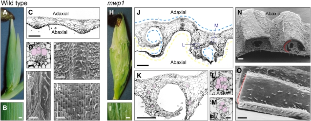
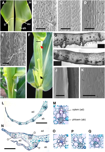
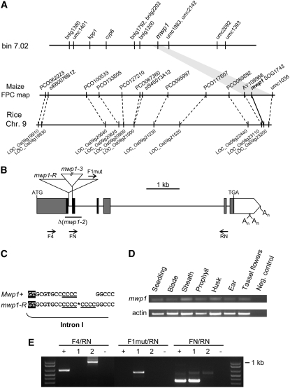
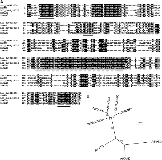
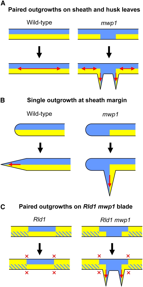
Leaf primordia initiate from the shoot apical meristem with inherent polarity; the adaxial side faces the meristem, while the abaxial side faces away from the meristem. Adaxial/abaxial polarity is thought to be necessary for laminar growth of leaves, as mutants lacking either adaxial or abaxial cell types often develop radially symmetric lateral organs. The milkweed pod1 (mwp1) mutant of maize (Zea mays) has adaxialized sectors in the sheath, the proximal part of the leaf. Ectopic leaf flaps develop where adaxial and abaxial cell types juxtapose. Ectopic expression of the HD-ZIPIII gene rolled leaf1 (rld1) correlates with the adaxialized regions. Cloning of mwp1 showed that it encodes a KANADI transcription factor. Double mutants of mwp1-R with a microRNA-resistant allele of rld1, Rld1-N1990, show a synergistic phenotype with polarity defects in sheath and blade and a failure to differentiate vascular and photosynthetic cell types in the adaxialized sectors. The sectored phenotype and timing of the defect suggest that mwp1 is required late in leaf development to maintain abaxial cell fate. The phenotype of mwp1; Rld1 double mutants shows that both genes are also required early in leaf development to delineate leaf margins as well as to initiate vascular and photosynthetic tissues.


Figures

Figure 1.
Figure 1.
Phenotype of Wild-Type and mwp1-R Husk Leaves. (A) to (G) Wild-type husk leaves. (A) Wild-type ear covered by smooth husk leaves. (B) Magnified view of the surface of a wild-type husk leaf. (C) Transverse section through a wild-type husk leaf. (D) Close-up view of a vascular bundle, showing a collateral distribution of adaxial xylem (pseudocolored pink) and abaxial phloem. (E) Scanning electron micrograph of the abaxial epidermis. (F) Scanning electron micrograph of the abaxial epidermis near the margin, showing characteristically long hairs. (G) Scanning electron micrograph of the adaxial epidermis. (H) to (O) mwp1-R husk leaves. (H) mwp1-R ears are covered by rough husk leaves with outgrowths. (I) Magnified view of the surface of a mwp1-R husk leaf. (J) Transverse section through a mwp1-R husk leaf, showing the development of pairs of flaps flanking sectors of adaxialized epidermis on the abaxial side. Parts of the epidermis with adaxial and abaxial characteristics are indicated by blue and yellow lines, respectively. (K) Magnified view of (J), showing that the vascular bundles in the flaps have xylem oriented toward the inner, adaxialized side. (L) and (M) Examples of radialized bundles from mwp1-R leaf flaps, with xylem at the periphery (pseudocolored pink). (N) Scanning electron micrograph of a piece of mwp1-R husk leaf, showing the hairy outer surface of the flaps, characteristic of abaxial margin identity. (O) Scanning electron micrograph of the inner surface of a flap, showing adaxial sheath characteristics. Bars = 1 mm in (B) and (I), 20 μm in (D), (L), and (M), and 200 μm in all others.
Figure 2.
Figure 2.
The mwp1 Phenotype in Vegetative Leaves. (A) Wild-type leaves consist of three domains along the proximal/distal axis: sheath (sh), auricle (au), and blade (bl). The ligule (li) normally develops at the sheath-auricle boundary on the adaxial side (left) and is absent from the abaxial side (right). (B) to (E) Different domains of the wild-type leaf are distinguishable based on epidermal characteristics. (B) Adaxial blade, showing macrohairs. (C) Abaxial blade, lacking macrohairs. (D) Adaxial sheath, which is not hairy. (E) Abaxial sheath, which is hairy. (F) mwp1-R mutants have an ectopic ligule at the sheath-auricle boundary as well as adaxialized sectors (red arrow) that extend below the ligule and above the node, sometimes with development of large laminar flaps (black arrow). The horizontal line marks the position of the section shown in (G). (G) Transverse section through a mwp1 mutant sheath, showing ectopic cell proliferation and extra vascular bundles lacking the abaxial hypodermal sclerenchyma. (H) Transverse section through the wild-type sheath. (I) Single flaps (white arrow) often develop at the sheath margins. (J) Scanning electron micrograph of the margin region, including a nonhairy margin flap (left) that develops adjacent to normal-looking hairy sheath (right). (K) Close-up view of the flap epidermis showing absence of hairs. (L) Transverse section across the tapered sheath margin of a wild-type leaf. (M) Close-up view of a wild-type vascular bundle, showing adaxial (ad) xylem and abaxial (ab) phloem. (N) Transverse section through the margin region of a mwp1-R mutant leaf, showing the true margin (blunt) and the ectopic margin (tapered). (O) to (Q) Close-up views of mwp1-R vascular bundles, with xylem at a peripheral location, as in (O) and (Q), or correctly oriented, as in (P). Bars = 500 μm in (E), 1 mm in (J), and 200 μm in all others.
Figure 3.
Figure 3.
Molecular Cloning and Characterization of mwp1 Alleles. (A) Mapping strategy used to clone mwp1. mwp1 was first mapped to bin 7.02 using molecular markers and subsequently assigned to an interval defined by markers AY109968 and umc1036 on a contig of the physical map with synteny to rice chromosome 9. (B) Structure of the mwp1 transcriptional unit, which consists of six exons (boxes) that encode an open reading frame (shaded boxes) with similarity to members of the KAN protein family. The sequence encoding the GARP domain characteristic of these proteins is in black. Triangles mark the insertions in the mwp1-R and mwp1-3 alleles. The region deleted in the mwp1-2 allele is indicated by a horizontal line. Arrows (not to scale) indicate the approximate position of some of the oligonucleotides used. An, polyadenylation sites. (C) Duplication of four nucleotides at the insertion site of a retrotransposon in mwp1-R. A retrotransposon was found inserted at the site marked by an asterisk, 13 nucleotides downstream of the exon/intron boundary, and was flanked by a direct repeat of four nucleotides (underlined). The first two nucleotides (GT) of intron I are highlighted. (D) RT-PCR analysis of mwp1 expression in assorted tissues. (E) RT-PCR analysis of mwp1 expression in wild-type and mutant seedlings. Primers F4 and RN fail to amplify a band using cDNA from mwp1-R mutant seedlings but amplify larger products in mwp1-2 due to mis-splicing. Primers F1-mut (specific to the mwp1-R insertion) and RN amplify a processed chimeric transcript only in mwp1-R, consisting of retrotransposon and gene sequences. Primers FN (which is specific to the region deleted in the mwp1-2 allele) and RN amplify bands of the expected size using cDNA of wild-type and mwp1-R seedlings. Lanes correspond to the wild type (+), mwp1-R (1), mwp1-2 (2), and water control (−). Molecular weight marker is 1 kb Plus (Invitrogen).
Figure 4.
Figure 4.
Sequence Alignment and Phylogenetic Analysis of mwp1 and Related KAN Genes. (A) Alignment of the amino acid sequences of mwp1 and the four most similar proteins in maize and rice. A dashed line indicates the GARP domain. Black lines indicate additional conserved regions in KAN proteins, first identified by Iwasaki and Nitasaka (2006), which are also conserved in KAN proteins from grasses. (B) Neighbor-joining phylogenetic tree based on an alignment of the nucleotide sequence coding for the GARP domain. Numbers at the branches are percentages based on 10,000 bootstrap repetitions.
Figure 5.
Figure 5.
In Situ Hybridization Analysis of mwp1 Expression. (A) to (C) Transverse sections of young seedlings 2 d after germination, showing several developing leaves covered by the coleoptile (CO). The oldest (most external) leaf primordia are at plastochron 6 (P6) stage and were sectioned at the level of the sheath. (A) Wild-type seedling. (B) mwp1-2 seedling. (C) Rld1-N1990 seedling. (D) Magnified view of the sheath of a wild-type developing leaf, showing higher expression in the abaxial epidermis. (E) Magnified view of (C) showing reduced expression in the abaxial epidermis. (F) Close-up view of a wild-type vascular bundle showing mwp1 expression between the xylem and phloem. (G) Longitudinal section of a wild-type shoot apical meristem and the surrounding leaf primordia. (H) Close-up view of the meristem in (G). Expression is absent from the apex of the meristem. (I) Transverse section of a wild-type shoot apical meristem showing strong expression in the margins of P2 and P3 leaf primordia.
Figure 6.
Figure 6.
In Situ Hybridization Analysis of rld1 Expression in Wild-Type and mwp1 Plants. (A) and (B) Transverse sections of female axillary shoots, showing the developing ear (e) covered by several husk leaves (h) and a prophyll (p). (A) The wild type. (B) mwp1-R showing ectopic abaxial expression of rld1 in the husk leaves associated with developing pairs of leaf flaps. (C) and (D) Magnified views of the developing flap pairs in (B). The pairs of flaps in are indicated by red arrows. (E) Wild-type vegetative leaf margins showing adaxial rld1 expression. (F) mwp1-R vegetative leaves showing ectopic rld1 expression and a developing margin flap. (G) Magnified view of (F). Bars = 50 μm in (C) and (D), 500 μm in (F), and 200 μm in all others.
Figure 7.
Figure 7.
The Synergistic Phenotype of mwp1; Rld1 Double Mmutants. (A) Rld1-N1990/rld1+ mutant. (B) mwp1-R; Rld1-N1990/rld1+ double mutant showing longitudinal sectors that extend the length of sheath and blade. A leafless node is indicated by a white arrow. (C) Same genotype as in (B) but with an ectopic blade fused to and developing from a sector in the main leaf. (D) mwp1-R; Rld1-N1990/rld1+ double mutant leaves extended to show the shape and propagation of the sectors from sheath to blade. (E) Scanning electron micrograph of the sheath of a mwp1-R; Rld1-N1990/rld1+ mutant leaf, as seen from the abaxial side, showing normal-looking sheath (left side) and the adaxialized, hairless epidermis of a sector (right side). (F) Magnified view of the adaxialized epidermis of the sector shown in (E). (G) Sheath of mwp1-R; Rld1-N1990/rld1+ mutant leaf showing an adaxialized sector with small hairs (right side), in contrast with the hairy, normal-looking sheath of adjacent tissue. (H) Scanning electron micrograph of the sectors and flaps observed on the abaxial surface of mwp1-R; Rld1-N1990/rld1+ leaf blades. (I) Transverse section corresponding to the region shown in (H). Dashed lines connect equivalent positions in (H) and (I), corresponding to developing ectopic blade margins. (J) Transverse section of an mwp1-R leaf that, like the wild type, shows bulliform cells only on the adaxial epidermis (arrow). (K) Transverse section of a Rld1-N1990/rld1+ leaf, showing bulliform cells (arrow) on the abaxial side but not on the adaxial. (L) Detailed view of (I), showing abaxial bulliform cells (arrow) as in (K).
Figure 8.
Figure 8.
Model for the Development of Laminar Outgrowths in mwp1 Mutants and in mwp1; Rld1 Double Mutants. (A) In wild-type husk leaves, the juxtaposition of adaxial (blue) and abaxial (yellow) cell types promotes laminar growth (red arrows). In mwp1 leaves, a failure to initiate or maintain abaxial identity in husk leaves results in ectopic adaxial cell types on the adaxial side, creating new boundaries of juxtaposed abaxial and adaxial identity. As predicted by the juxtaposition model, growth then occurs at the planes defined by these new boundaries. (B) When ectopic adaxial cell types are present at the sheath margin, a single ectopic boundary is created, resulting in the development of a single outgrowth. (C) Rld1 mutations often cause a complete switch in the abaxial/adaxial polarity of the blade. Sectors with normal polarity are often located adjacent to sectors with reversed polarity, but this juxtaposition does not normally lead to laminar outgrowths (red crosses). Development of outgrowths in mwp1; Rld1 blades may occur in response to adaxialization in sectors of the blade that otherwise retain abaxial identity on the abaxial side.

References

    1. Bao, N., Lye, K.W., and Barton, M.K. (2004). MicroRNA binding sites in Arabidopsis class III HD-ZIP mRNAs are required for methylation of the template chromosome. Dev. Cell 7 653–662. - PubMed
    1. Barrero, J.M., Gonzalez-Bayon, R., del Pozo, J.C., Ponce, M.R., and Micol, J.L. (2007). INCURVATA2 encodes the catalytic subunit of DNA polymerase alpha and interacts with genes involved in chromatin-mediated cellular memory in Arabidopsis thaliana. Plant Cell 19 2822–2838. - PMC - PubMed
    1. Blauth, S.L., Yao, Y., Klucinec, J.D., Shannon, J.C., Thompson, D.B., and Guilitinan, M.J. (2001). Identification of Mutator insertional mutants of starch-branching enzyme 2a in corn. Plant Physiol. 125 1396–1405. - PMC - PubMed
    1. Bortiri, E., Chuck, G., Vollbrecht, E., Rocheford, T., Martienssen, R., and Hake, S. (2006). ramosa2 encodes a LATERAL ORGAN BOUNDARY domain protein that determines the fate of stem cells in branch meristems of maize. Plant Cell 18 574–585. - PMC - PubMed
    1. Edgar, R.C. (2004). MUSCLE: A multiple sequence alignment method with reduced time and space complexity. BMC Bioinformatics 5 1–19. - PMC - PubMed

Publication types

MeSH terms

LinkOut - more resources