. 2008 Aug;20(8):2146-59.
doi: 10.1105/tpc.107.056812. Epub 2008 Aug 29.

Arabidopsis TONNEAU1 proteins are essential for preprophase band formation and interact with centrin

Affiliations

Arabidopsis TONNEAU1 proteins are essential for preprophase band formation and interact with centrin

Juliette Azimzadeh et al. Plant Cell. 2008 Aug.

Abstract

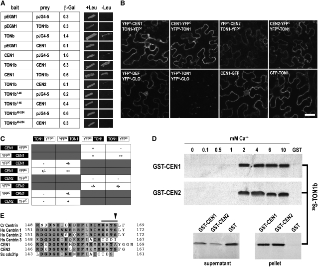
Plant cells have specific microtubule structures involved in cell division and elongation. The tonneau1 (ton1) mutant of Arabidopsis thaliana displays drastic defects in morphogenesis, positioning of division planes, and cellular organization. These are primarily caused by dysfunction of the cortical cytoskeleton and absence of the preprophase band of microtubules. Characterization of the ton1 insertional mutant reveals complex chromosomal rearrangements leading to simultaneous disruption of two highly similar genes in tandem, TON1a and TON1b. TON1 proteins are conserved in land plants and share sequence motifs with human centrosomal proteins. The TON1 protein associates with soluble and microsomal fractions of Arabidopsis cells, and a green fluorescent protein-TON1 fusion labels cortical cytoskeletal structures, including the preprophase band and the interphase cortical array. A yeast two-hybrid screen identified Arabidopsis centrin as a potential TON1 partner. This interaction was confirmed both in vitro and in plant cells. The similarity of TON1 with centrosomal proteins and its interaction with centrin, another key component of microtubule organizing centers, suggests that functions involved in the organization of microtubule arrays by the centrosome were conserved across the evolutionary divergence between plants and animals.


Figures

Figure 1.
Figure 1.
Developmental Phenotype of the ton1 Mutant. (A) ton1 mutant seeds (bottom) have a modified shape compared with the wild type (top). Bar = 1 mm. (B) In vitro–grown wild-type (left) and ton1 mutant plantlets 7 d after germination. Bar = 5 mm. (C) to (E) Wild-type plant (C) and ton1 mutants 11 d (D) and 3 weeks (E) after germination. Bars = 5 mm. (F) and (G) ton1 mutant plant 6 weeks after germination. Bars = 5 mm. (H) Close-up on a ton1 mutant flower. Bar = 1 mm. (I) From left to right: in vitro–grown ton1, fass2, fass11, and fass12 (= ton2-12; Camilleri et al., 2002) mutants 10 d after germination. ton1 and fass2 are in the Wassilewskija (Ws) background, while fass11 and 12 are in the Landsberg erecta (Ler) background. Bar = 5 mm.
Figure 2.
Figure 2.
MT Organization in the ton1 Mutant. (A) and (B) Interphase cortical arrays of MTs in wild-type (A) and ton1 (B) hypocotyl cells of transgenic Arabidopsis expressing GFP-MBD, which labels MTs. Arrows indicate the direction of the long axis of hypocotyls. (C) to (J) Immunolocalization in root cells using an anti-α-tubulin antibody. (C) A typical wild-type PPB of MTs just before transition toward spindle assembly. (D) Preprophase nuclei in dividing ton1 mutant cells, showing dense accumulation of MTs around the nuclei and no evidence of cortical structures. (E) Wild-type spindles. (F) A ton1 mutant spindle. (G) Wild-type phragmoplast. (H) ton1 mutant phragmoplast. (I) A wild-type Arabidopsis root tip. (J) A ton1 mutant root tip, showing alteration in cell shape, size, number, and loss of global cellular organization. Bars = 50 μm in (A), (B), (I), and (J) and 10 μm in (C) to (H).
Figure 3.
Figure 3.
Structure of the TON1 Locus. The TON1a gene contains eight exons (black boxes), whereas the TON1b gene has only seven exons (white boxes), as TON1a exons 6 and 7 are fused in TON1b. The 1.4-kb deletion found at the translocation breakpoint in the ACL4 line is boxed. Restriction sites corresponding to fragments used in complementation experiments are shown on top; their positions on Arabidopsis Genome Initiative pseudomolecules (version 5, ftp://ftp.Arabidopsis.org/home/tair/Sequences/) are: SalI 20,387,939 bp; PvuII 20,395,408; XhoI 20,398,868 bp.
Figure 4.
Figure 4.
Analysis of TON1a and TON1b Gene Expression by RT-PCR. RT-PCR analyses were performed as described (Camilleri et al., 2002) using gene-specific primers: a 461-bp RT-PCR fragment was obtained for the TON1a transcript using the primers Spec1aF and Spec1aR, specific for the TON1a gene, and a 281-bp RT-PCR fragment was obtained for TON1b using the primers Spec1bF and Spec1bR, specific for the TON1b gene. The constitutively expressed APT1 gene (Moffatt et al., 1994) was used as a control (564-bp RT-PCR fragment with primers APT-RT1 and APT-RT2). WS gDNA, Ws genomic DNA; No RT, negative control (no reverse transcriptase). In the ton1 mutant, primers specific for the TON1b gene detect a fusion transcript (asterisks) between (1) the Basta resistance gene present in the T-DNA used for insertion mutagenesis and (2) the 3′ end of the TON1b gene.
Figure 5.
Figure 5.
Multiple Alignment of Proteins Related to TON1 Defines a New Conserved Protein Motif. (A) The N termini of several proteins belonging to various taxonomic groups were aligned. For clarity, only one representative sequence of each taxonomic group is shown here. Similarities are boxed, identities are in dark gray, and similarities are in light gray. Three highly conserved regions appear from the alignment: the TOF motif is located at the very beginning of all protein sequences. The second is a LisH dimerization motif (Emes and Ponting, 2001). A third region with a conserved PLL triad is presumably involved in LisH-mediated dimerization. Arrowheads indicate positions of deleterious mutations characterized in the human OFD1 gene (Romio et al., 2003). (B) Phylogenetic tree of TON1 homologs. Multiple alignments of the ∼180 N-terminal residues of each sequence were computed with Clustal, MAFFT, and Muscle algorithms (Thompson et al., 1994; Katoh et al., 2002; Edgar, 2004) and hand-curated to produce a consensus alignment. The alignment was used to produce an unrooted UPGMA tree. Bootstrap values are indicated on main branches (1000 repetitions). Four groups emerge from the N-terminal sequence alignment: land plant TON1, vertebrate FOP, vertebrate OFD, and a group of short (∼175 residues) proteins related to FOP from various eukaryotes (FOP-like). The Chlamydomonas FOP is equally distant to TON1 and FOP. At, Arabidopsis thaliana; Os, Oryza sativa; Pi, Pinus tadea; Cy, Cycas rumphii; Pp, Physcomitrella patens; Hs, Homo sapiens; Gg, Gallus gallus; Xl, Xenopus laevis; Dr, Danio rerio; Ci, Ciona Intestinalis; Cr, Chlamydomonas reinhardtii; Tn, Tetraodon nigroviridis; Nv, Nematostella vectensis; Pt, Paramecium tetraurelia; Lm, Leishmania majo; Tb, Trypanosoma brucei; Am, Apis mellifera; Tv, Trichomonas vaginalis; Gl, Giardia lamblia. Accession numbers are listed in Supplemental Figure 4 online. (C) Comparison of TON1 and FOP reveals a similar organization at their N terminus, with conserved motifs described above, a short Ser-rich region (S), and Ser phosphorylation sites (Benschop et al., 2007) around positions 150 to 160. FOP also has a Tyr phosphorylation site at position 337 (http://www.phosphosite.org/).
Figure 6.
Figure 6.
Subcellular Localization of the TON1 Proteins. Top panel: Protein gel blot analysis of total proteins of ton1 and wild-type 10-d-old in vitro–grown seedlings probed against the anti-TON1 polyclonal serum and an anti-α-tubulin antibody. TON1 proteins are undetectable in the ton1 mutant extract. Bottom panel: Total proteins and proteins from soluble (S) and pellet (P) fractions obtained from adult rosette leaves were separated by electrophoresis, transferred to membrane, and probed with anti-TON1 serum, anti-α-tubulin antibody, and anti-H+-ATPase serum. Upon exposure to high pH (pH 11.5), TON1 proteins were released from the pellet fraction and solubilized, whereas resuspension of the pellet in the homogenization buffer used to make the original protein extract did not. As a control, the intrinsic membrane protein H+-ATPase is not released from membranes after exposure to high pH. Size of the proteins is as follows: TON1, 30 kD; tubulin, 50 kD; H+-ATPase, 100 kD.
Figure 7.
Figure 7.
Localization of a GFP-TON1 Fusion in Arabidopsis and Tobacco Cells. (A) and (B) In the Arabidopsis root tip of 5-d-old seedlings, in addition to a cytoplasmic staining, the GFP-TON1a fusion faintly but consistently labels the PPB (asterisk in [A] and cell in [B]). In actively dividing cell files, GFP-TON1a also accumulates on the transverse sides of cells (A). (C) and (D) The same pattern is observed in transformed tobacco roots expressing GFP-TON1a; see labeled cross-wall in (C) and two cells displaying PPB in (D). (E) and (F) In Arabidopsis expanding hypocotyl cells of 3-d-old etiolated seedlings, GFP-TON1a accumulates as discrete punctate staining organized into dense linear arrays, reminiscent of MT organization in expanding cells. Usually, most of expanding hypocotyls cells display MT arrays roughly transverse to the cell axis similar to the GFP-TON1a staining seen in the left cell in (E) and in (F), although some cells can have more longitudinal arrays as the right cell in (E) (similar patterns are described in Paredez et al., 2006; Chan et al., 2007). (G) to (J) Arabidopsis hypocotyl cells expressing GFP-TON1a before (G) and after incubation in 10 μM oryzalin for 5 min (H), 15 min (I), and 70 min (J). Micrographs in (A) to (D) are confocal laser scanning microscope images, and images in (E) to (J) were obtained using a spinning disk confocal microscope. All plantlets express the GFP-TON1a fusion under the control of the 35S promoter. Bars = 20 μm in (A), (C), and (D) to (F) and 10 μm in (B) and (G) to (J).
Figure 8.
Figure 8.
Interaction of TON1 with Arabidopsis Centrins. (A) Yeast two-hybrid experiments. Bait TON1b fragments and CEN1 were cloned as LexA fusions; pEGM1 and pJG4-5 are empty vector controls. Full-length TON1b, CEN1, and CEN2 prey fragments were cloned in pJG4-5. β-Gal activity is expressed in arbitrary units. Left panels: Growth of yeast cells on nonselective medium with Leu (+Leu). Right panels: Growth of yeast clones on selective medium without Leu (−Leu). The LEU2 reporter in EGY48 is known to be more sensitive than the β-Gal reporter for some baits (Ausubel et al., 1998), which may explain the low level of β-Gal for CEN1-TON1b compared with its growth on −Leu medium. (B) BiFC visualization of TON1a interaction with Arabidopsis centrins. Shown are confocal images of tobacco epidermal cells coinfiltrated with Agrobacterium cultures harboring expression vectors. In the typical jigsaw puzzle cells, the cytoplasm and nucleus are restricted to the cell's periphery by the large central vacuole. When transiently overexpressed under the control of the 35S promoter, CEN1-GFP localizes to the cytoplasm and the nucleus (bottom panel, middle right), and GFP-TON1 localizes to the cytoplasm (bottom panel, right). Positive control for the BiFC experiment corresponds to coexpression of YFPN-DEF and YFPC-GLO (bottom panel, left), the documented heterodimeric complex formation between Anthirrinum majus MADS box transcription factors DEFICIENS and GLOBOSA (Schwarz-Sommer et al., 1992). Negative controls correspond to expression of the protein of interest (TON1 or CEN1 or 2) fused to YFP moiety alone or to coexpression of these protein fusions with unrelated proteins (i.e., DEFICIENS and GLOBOSA). Here, coexpression of YFPN-TON1 and YFPC-GLO is shown (bottom panel, middle left). Top panels correspond to examples of YFP complementation obtained after coexpression of the indicated fusion proteins. As expected in these conditions of strong overexpression, the location of TON1-centrin complexes formation is cytoplasmic. Bar = 25 μm. (C) Summary of interactions between TON1a and Arabidopsis centrins, CEN1 and CEN2, assayed by BiFC in epidermal cells of N. benthamiana. ++, strong fluorescent signal; +, significant fluorescent ± weak signal above background; −, no significant signal. (D) In vitro pull-down assays. CEN1 and CEN2 were produced in E. coli as fusions with the GST glutathione binding domain. 35S-TON1b was produced in vitro in a coupled transcription/translation system. Top panels: The GST fusions were bound on gluthathione-agarose, washed and incubated with 35S-TON1b in the presence of increasing concentrations of Ca2+ (from 0 to 10 mM), before elution and analysis by SDS-PAGE and autoradiography. GST, negative control (GST alone, 10 mM Ca2+). Bottom panels: Experiment showing the supernatant (left) and pellet (right) of a 35S-TON1b pull-down assay with GST-CEN1, GST-CEN2, and GST alone under 10 mM Ca2+. (E) Amino acid alignment of the C-terminal part of Chlamydomonas reinhardtii centrin, human centrins 1, 2, and 3, Arabidopsis CEN1 and CEN2, and yeast cdc31p, showing differences at a C-terminal phosphorylation site for cAMP-dependent protein kinase ([R/K]-[R/K]-x-[S/T]; bar) and presence of a phosphorylatable Ser (arrowhead). Similarities are boxed, and identities are in bold.

References

    1. Andersen, J.S., Wilkinson, C.J., Mayor, T., Mortensen, P., Nigg, E.A., and Mann, M. (2003). Proteomic characterization of the human centrosome by protein correlation profiling. Nature 426 570–574. - PubMed
    1. Ausubel, F.M., Brent, R., Kingston, R.E., Moore, D.D., Seidman, J.G., Smith, J.A., and Struhl, K. (1998). Current Protocols in Molecular Biology. (New York: John Wiley & Sons).
    1. Ayaydin, F., Vissi, E., Meszaros, T., Miskolczi, P., Kovacs, I., Feher, A., Dombradi, V., Erdodi, F., Gergely, P., and Dudits, D. (2000). Inhibition of serine/threonine-specific protein phosphatases causes premature activation of cdc2MsF kinase at G2/M transition and early mitotic microtubule organisation in alfalfa. Plant J. 23 85–96. - PubMed
    1. Baskin, T.I., Meekes, H.T.H.M., Liang, B.M., and Sharp, R.E. (1999). Regulation of growth anisotropy in well-watered and water-stressed maize roots. II. Role of cortical microtubules and cellulose microfibrils. Plant Physiol. 119 681–692. - PMC - PubMed
    1. Bechtold, N., Ellis, J., and Pelletier, G. (1993). In planta Agrobacterium mediated gene transfer by infiltration of adult Arabidopsis thaliana plants. C. R. Acad. Sci. III 316 1194–1199.

Publication types

MeSH terms

Substances

Associated data