Review
. 2009 Feb 12;113(7):1399-407.
doi: 10.1182/blood-2008-07-019307. Epub 2008 Aug 29.

TLRs and innate immunity

Affiliations
Review

TLRs and innate immunity

Bruce A Beutler. Blood. .

Abstract

One of the most fundamental questions in immunology pertains to the recognition of non-self, which for the most part means microbes. How do we initially realize that we have been inoculated with microbes, and how is the immune response ignited? Genetic studies have made important inroads into this question during the past decade, and we now know that in mammals, a relatively small number of receptors operate to detect signature molecules that herald infection. One or more of these signature molecules are displayed by almost all microbes. These receptors and the signals they initiate have been studied in depth by random germline mutagenesis and positional cloning (forward genetics). Herein is a concise description of what has been learned about the Toll-like receptors, which play an essential part in the perception of microbes and shape the complex host responses that occur during infection.


Figures
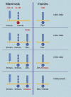
Figure 1
Figure 1
Key developments in the TLR field over time. Conceptual advances shown in red for both insects and mammals. Toll was identified as a developmental protein in 1985. CD14 was identified as a part of the LPS receptor in 1990; the IL-1 receptor was cloned in 1988 and noted to have domain homology to Toll in 1991. In 1994, mammalian TLRs were first identified, but incorrectly assumed to have developmental functions based on what was known in Drosophila at the time. In 1996, the dual immunologic/developmental character of Drosophila Toll was recognized. The immune function of a mammalian TLR was first demonstrated in 1998. Illustration made with assistance of Marie Dauenheimer.
Figure 2
Figure 2
Homologies between mammalian and insect immune defense pathways. In mammals (enclosed in dashed curve), TLR and TNF signaling pathways are unified, in that TNF is produced in response to all TLR ligands, and then signals via the TNF receptors to induce cell death and NF-κB activation in other cells. In Drosophila, the Toll pathway and the Imd pathway are separated, each responding independently to microbial stimuli. Molecular homologies are shaded in blue. AMP indicates antimicrobial peptide. Genes, implies the induction of many hundreds of NF-κB dependent genes. Figure is not all encompassing and is meant to emphasize core similarities between the pathways. Illustration made with assistance of Marie Dauenheimer.
Figure 3
Figure 3
Inbreeding protocol for generating homozygous mutations. G0 mice are bred to germline mutant G1 females, and siblings from both the G1 and G2 generations are crossed. The chance for homozygosity for each G1 mutation is only 1 in 16 in every G3 mouse. Red asterisks indicate mutations originating from the G0 sire; blue asterisks indicate mutations originating in the G1 dam. Star size indicates derivation from an immediate ancestor (large) or from a more remote ancestor (small). Generations (yellow boxes) are aligned. This strategy has the advantage of introducing X-linked mutations into the pedigree, and causes homozygosity for autosomal mutations at a rate 1.39 times greater than would be the case if additional mutations were not introduced by breeding each G0 with a G1 mutant female. Illustration made with assistance of Marie Dauenheimer.
Figure 4
Figure 4
The ectodomain of a TLR3 subunit. “Worms” rendering with yellow arrows indicating beta sheet and green coil indicating alpha helix, imaged with the program CN3D. From NCBI protein database, PDB 1ZIW. Amino terminal (N) and carboxy terminal (c) ends of the structure are indicated. Loops outside the solenoid (arrow) may give flexibility to the protein. Carbohydrate residues (ball and stick) may influence association between subunits and/or binding of ligand.
Figure 5
Figure 5
TLR signaling pathways established by mutagenesis. Each red × represents a different mutation. The relationship of proteins within the signaling pathways has been deduced biochemically and by reference to domain structures of the target molecules. Some mutations have yet to be found, but affect proteins critical for signal transduction. Illustration made with assistance of Marie Dauenheimer.

Similar articles

Cited by

References

    1. Janeway CA., Jr Approaching the asymptote? Evolution and revolution in immunology 20. Cold Spring Harb Symp Quant Biol. 1989;54(Pt 1):1–13. - PubMed
    1. Pfeiffer R. Untersuchungen über das Choleragift. Z Hygiene. 1892;11:393–412.
    1. Shear MJ, Andervont HB. Chemical treatment of tumors. III. Separation of hemorrhage-producing fraction of B. coli filtrate. Proc Soc Exp Biol Med. 1936;34:323–325.
    1. Hartwell JL, Shear MJ, Adams JR., Jr Chemical treatment of tumors. VII. Nature of the hemorrhage-producing fraction from Serratia marcescens (Bacillus prodigiosus) culture filtrate. J Natl Cancer Inst. 1943;4:107–122.
    1. Kahler H, Shear MJ, Hartwell JL. Chemical treatment of tumors. VIII. Ultracentrifugal and electrophoretic analysis of the hemorrhage-producing fraction from Serratia marcescens (Bacillus prodigiosus) culture filtrate. J. Natl Cancer Inst. 1943;4:123–129.

Publication types

Substances