Review
. 2008 Nov;49(11):2283-301.
doi: 10.1194/jlr.R800018-JLR200. Epub 2008 Aug 29.

Thematic review series: glycerolipids. DGAT enzymes and triacylglycerol biosynthesis

Affiliations
Review

Thematic review series: glycerolipids. DGAT enzymes and triacylglycerol biosynthesis

Chi-Liang Eric Yen et al. J Lipid Res. 2008 Nov.

Abstract

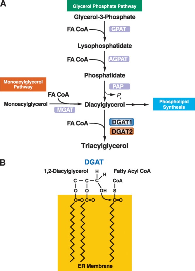
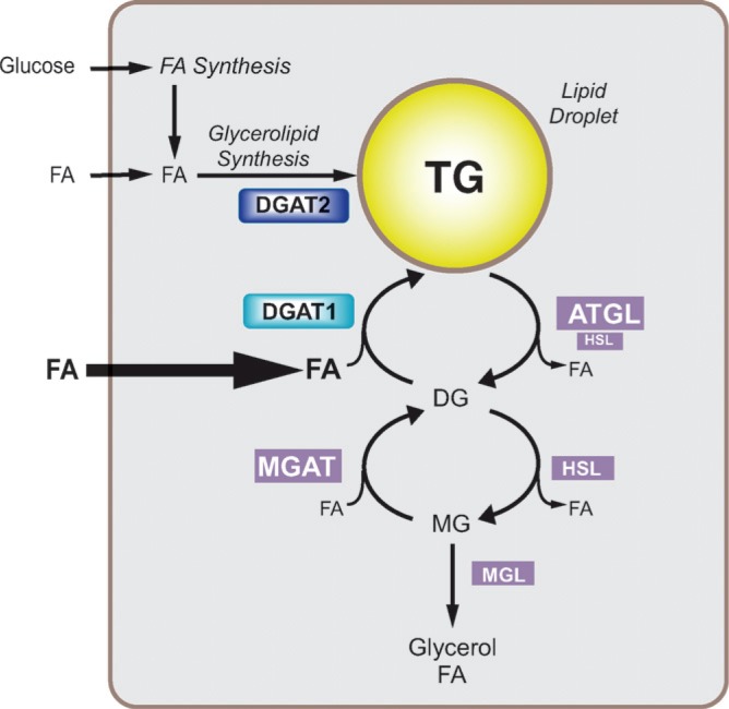
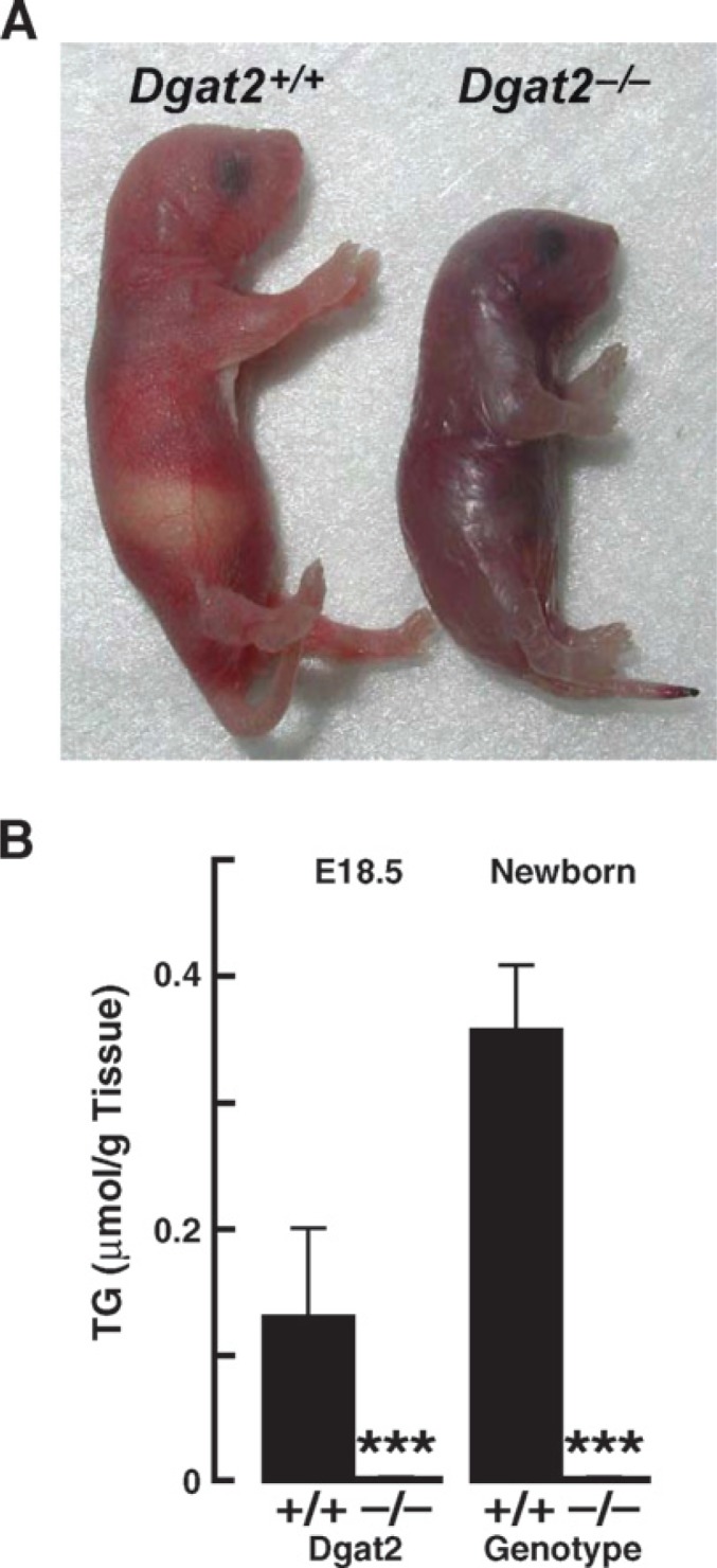
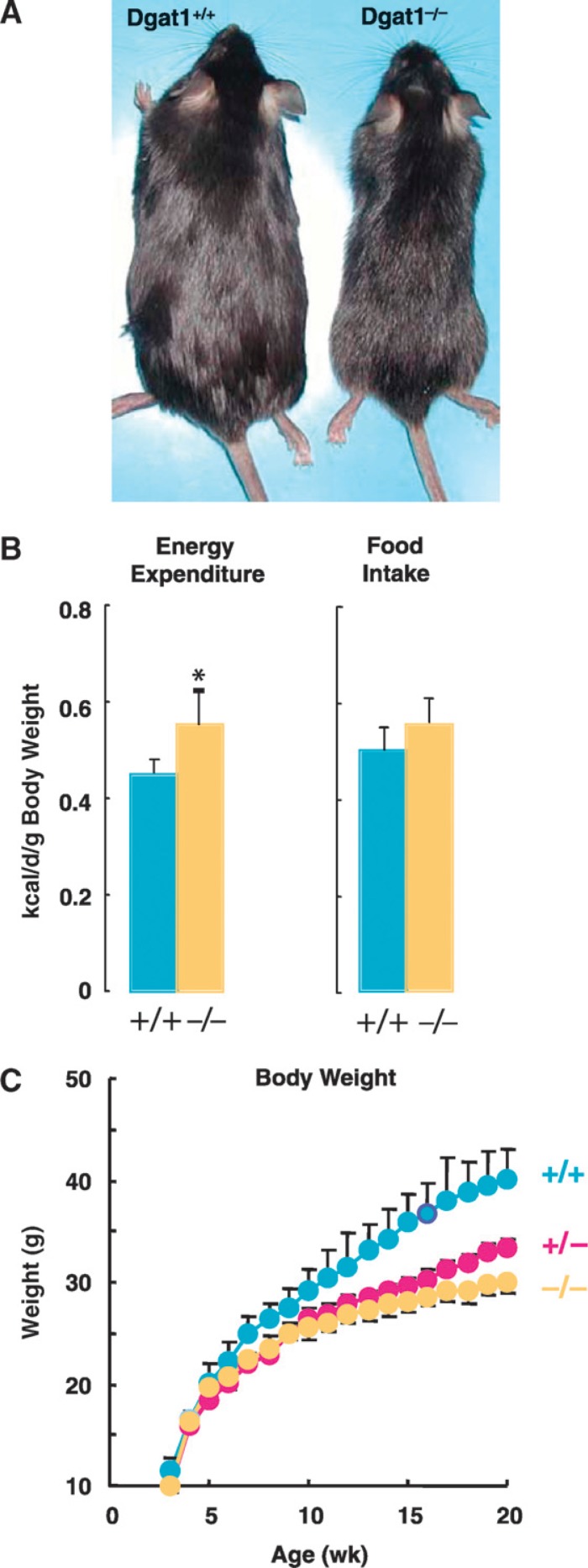
Triacylglycerols (triglycerides) (TGs) are the major storage molecules of metabolic energy and FAs in most living organisms. Excessive accumulation of TGs, however, is associated with human diseases, such as obesity, diabetes mellitus, and steatohepatitis. The final and the only committed step in the biosynthesis of TGs is catalyzed by acyl-CoA:diacylglycerol acyltransferase (DGAT) enzymes. The genes encoding two DGAT enzymes, DGAT1 and DGAT2, were identified in the past decade, and the use of molecular tools, including mice deficient in either enzyme, has shed light on their functions. Although DGAT enzymes are involved in TG synthesis, they have distinct protein sequences and differ in their biochemical, cellular, and physiological functions. Both enzymes may be useful as therapeutic targets for diseases. Here we review the current knowledge of DGAT enzymes, focusing on new advances since the cloning of their genes, including possible roles in human health and diseases.


Figures

Fig. 1
Fig. 1
Triacylglycerol synthesis and acyl-CoA:diacylglycerol acyltransferase (DGAT) enzymes. A: Triacylglycerols (triglycerides) are the end-product of a multi-step pathway. DGAT1 or DGAT2 catalyzes the final reaction. B: DGAT enzymes catalyze the formation of an ester linkage between a fatty acyl CoA and the free hydroxyl group of diacylglycerol. The model shows this reaction occurring at the surface of the endoplasmic reticulum (ER) bilayer membrane. GPAT, glycerol-phosphate acyltransferase; AGPAT, acylglycerol-phosphate acyltransferase; PAP, phosphatidic acid phosphohydrolase; MGAT, acyl CoA:monoacylglycerol acyltransferase.
Fig. 2
Fig. 2
Hypothetical model illustrating the role of DGAT enzymes in triacylglycerol synthesis in the ER. Triacylglycerol products of the DGAT reaction may be channeled into the cores of cytosolic lipid droplets or triacylglycerol-rich lipoproteins for secretion in cells such as enterocytes and hepatocytes. Although the model depicts the reaction at the cytosolic surface of the ER, it has been proposed that the reaction may also take place at the luminal surface (see text for discussion). DGAT1 is shown as a homotetramer (59).
Fig. 3
Fig. 3
Dendrograms of the DGAT gene families. A: The acyl-CoA: cholesterol acyltransferase (ACAT)/DGAT1 gene family is part of a membrane-bound O-acyltransferase (MBOAT) superfamily of enzymes. Selected members shown are human proteins unless indicated. MBOAT1 and MBOAT2 are family members whose activities are unknown. Dendrograms were generated by ClustalW using Biology Workbench (http://workbench.sdsc.edu/). B: The DGAT2 gene family. DC (DGAT candidate) 3 refers to a family member whose activity is unknown.
Fig. 4
Fig. 4
Schematic representations of murine DGAT1 and DGAT2 proteins. Possible domains and sites of modification are shown. Transmembrane domains were predicted by the program SOSUI (http://bp.nuap.nagoya-u.ac.jp/sosui/sosui_submit.html). The two transmembrane domains in DGAT2 have been confirmed experimentally (47). Phosphorylation sites were predicted by PROSEARCH (http://workbench.sdsc.edu/). PKC, PKA, and Tyr represent potential phosphorylation sites for protein kinase C, protein kinase A, and tyrosine kinase, respectively.
Fig. 5
Fig. 5
Hypothetical model for intracellular roles of DGAT1 and DGAT2. On the basis of the available evidence, we speculate that DGAT2 function is closely linked to endogenous FA synthesis and esterification, whereas DGAT1 may be involved in the recycling of hydrolyzed triacylglycerols by reesterifying the FAs. DGAT1 may also play an important role in protecting cells from high concentrations of FAs. The model suggests that DGAT2 is most active with low substrate concentrations (e.g., with de novo FA synthesis) and that DGAT1 is most active with high substrate concentrations, such as with exogenous FA or high levels of lipolysis. FAs need to be activated by forming FA CoA (not shown). HSL, hormone-sensitive lipase; ATGL, adipose-tissue triglyceride lipase; MGL, monoacylglycerol lipase. Permission to use Fig. 5 granted by Elsevier Ltd.
Fig. 6
Fig. 6
Phenotype of DGAT2 deficiency in mice. A: Mice lacking DGAT2 are smaller, do not feed well, have abnormal skin, and die early in the postnatal period. B: Reduction in carcass triacylglycerols in Dgat2−/− mice. A and B, reproduced from (46).
Fig. 7
Fig. 7
Effects of DGAT1 deficiency on energy metabolism in mice. A: Male mice (10 weeks old) fed a high-fat diet for 2 weeks. Note the leaner appearance of the Dgat1−/− mouse. Image courtesy of C. Villanueva. B: Energy expenditure and food intake in wild-type and Dgat1−/− mice. C: Weight curves for inbred wild-type, Dgat1+/−, and Dgat1−/− mice fed a high-fat diet (unpublished observations).

References

    1. Stryer L. Biochemistry. W.H. Freeman & Co.; New York, NY: 1988. p. 471.
    1. Unger RH. Lipotoxic diseases. Annu. Rev. Med. 2002;53:319–336. - PubMed
    1. Friedman J. Fat in all the wrong places. Nature. 2002;415:268–269. - PubMed
    1. Kennedy E. Metabolism of lipides. Annu. Rev. Biochem. 1957;26:119–148. - PubMed
    1. Bell RM, Coleman RA. Enzymes of glycerolipid synthesis in eukaryotes. Annu. Rev. Biochem. 1980;49:459–487. - PubMed

Publication types

MeSH terms