. 2008 Sep;180(1):165-78.
doi: 10.1534/genetics.108.090423. Epub 2008 Aug 30.

A random mutagenesis approach to isolate dominant-negative yeast sec1 mutants reveals a functional role for domain 3a in yeast and mammalian Sec1/Munc18 proteins

Affiliations

A random mutagenesis approach to isolate dominant-negative yeast sec1 mutants reveals a functional role for domain 3a in yeast and mammalian Sec1/Munc18 proteins

Alan Boyd et al. Genetics. 2008 Sep.

Abstract

SNAP receptor (SNARE) and Sec1/Munc18 (SM) proteins are required for all intracellular membrane fusion events. SNAREs are widely believed to drive the fusion process, but the function of SM proteins remains unclear. To shed light on this, we screened for dominant-negative mutants of yeast Sec1 by random mutagenesis of a GAL1-regulated SEC1 plasmid. Mutants were identified on the basis of galactose-inducible growth arrest and inhibition of invertase secretion. This effect of dominant-negative sec1 was suppressed by overexpression of the vesicle (v)-SNAREs, Snc1 and Snc2, but not the target (t)-SNAREs, Sec9 and Sso2. The mutations isolated in Sec1 clustered in a hotspot within domain 3a, with F361 mutated in four different mutants. To test if this region was generally involved in SM protein function, the F361-equivalent residue in mammalian Munc18-1 (Y337) was mutated. Overexpression of the Munc18-1 Y337L mutant in bovine chromaffin cells inhibited the release kinetics of individual exocytosis events. The Y337L mutation impaired binding of Munc18-1 to the neuronal SNARE complex, but did not affect its binary interaction with syntaxin1a. Taken together, these data suggest that domain 3a of SM proteins has a functionally important role in membrane fusion. Furthermore, this approach of screening for dominant-negative mutants in yeast may be useful for other conserved proteins, to identify functionally important domains in their mammalian homologs.


Figures

F<sc>igure</sc> 1.—
Figure 1.—
Mapping of dominant-negative mutations in SEC1. (A) The open reading frame of three strong dominant-negative mutants (D9, D18, and D25) was fully sequenced, revealing a clustering of mutations (represented by solid circles) in a central region of the SEC1 gene. Further sequencing showed that all but one of the remaining mutants contained a mutation in this region, corresponding to amino acids 328–370 (enclosed by rectangle). The amino acid sequence of this region is shown, with the various substitution mutations in boldface type. (B) To determine which of the multiple mutations in D18 and D25 were responsible for the dominant-negative phenotype, the SEC1 gene was divided into three restriction fragment cassettes: A, B, and C. These cassettes were then used to create domain-swap constructs carrying hybrid wild-type and mutant cassettes. The ability of the hybrids to confer the dominant-negative phenotype was then assessed by scoring growth inhibition on galactose media.
F<sc>igure</sc> 2.—
Figure 2.—
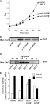
Effects upon growth and secretion of high-level expression of mutant sec1 genes. Wild-type cells were transformed with plasmids encoding wild-type (SEC1+) or dominant-negative (sec1-D18B+, sec1-D25B+) SEC1 constructs or with empty vector. Raffinose-grown transformants were reinoculated into fresh raffinose medium and pregrown before addition of galactose (2%: time = 0). Subsequent growth of the four strains was followed. After 360–400 min, samples were withdrawn for protein extraction and for assay of internal and external levels of invertase. (A) Growth of the four strains was followed by measurement of culture absorbance at 600 nm. (B) Detergent-soluble protein extracts of each strain were prepared and then analyzed by SDS–PAGE with subsequent immunoblotting using an anti-Sec1 antiserum. Note that the antibody fails to detect endogenous levels of Sec1 protein in control (vector-transformed) wild-type cells. (C) Different amounts of extracts from control (vector-transformed) and Sec1-overexpressing cells were immunoblotted with anti-Sec1 antiserum to calibrate the relative level of Sec1 protein overexpression. (D) Samples of cells and of culture medium from the various strains were assayed for the level of invertase activity. Open bars indicate intracellular invertase activity, and solid bars represent extracellular (secreted) invertase activity.
F<sc>igure</sc> 3.—
Figure 3.—
Overexpression of Sec9p, Sso2p, and Snc2p by high-copy plasmids. Wild-type cells were transformed with empty vector or plasmids carrying SEC9, SSO2, and SNC2 genes. These were then grown in liquid culture and detergent-solubilized protein extracts were prepared. The resulting samples were analyzed by SDS–PAGE and immunoblotting.
F<sc>igure</sc> 4.—
Figure 4.—
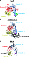
Dominant-negative sec1 mutations cluster in a structurally conserved domain in SM proteins. (A) The structure of Sec1 as predicted by I-TASSER computer simulations is shown with the dominant-negative mutations isolated in domain 3a highlighted (green, found in a single mutant; magenta, F361, mutated in four separate mutants). (B) The crystal structure of Munc18-1 bound to syntaxin 1a (pdb entry: 1dn1) is shown with the F361 equivalent residue, Y337, highlighted in magenta. (C) The crystal structure of Sly1 bound to Sed5 (pbd entry: 1mqs) is shown with the F361 equivalent residue, L390, highlighted in magenta. Structures were rendered using Chimera software.
F<sc>igure</sc> 5.—
Figure 5.—
The sec1 dominant-negative mutation equivalent in Munc18-1 (Y337L) slows exocytosis release kinetics. (A) Typical amperometric responses from untransfected cells (left) or cells transfected with the Munc18-1 Y337L mutant (right) following addition of digitonin and Ca2+ to elicit exocytosis. (B) Representative amperometric spikes from untransfected (left) or Y337L-transfected cells. (C) Analysis of the frequency of exocytotic fusion events reveals no significant difference between control and transfected cells. (D–F) Expression of Munc18-1 Y337L increases charge (C) and slows the kinetics of release by increasing both the rise time (D) and fall time (E) of individual amperometric spikes. (G) Representative example of a prespike foot (marked by arrow). (H–J) Analysis of the frequency (H), charge (I), and duration (J) of prespike feet reveals no significant difference between control and transfected cells. Data were analyzed from 240 spikes and 34 feet from 18 cells (control) and from 366 spikes and 100 feet from 26 cells (Y337L), and statistical significance was assessed using Mann–Whitney tests.
F<sc>igure</sc> 6.—
Figure 6.—
The Y337L mutation impairs binding of Munc18-1 to the neuronal SNARE complex, but not to syntaxin 1a. (A) 35S-radiolabeled, in vitro translated Munc18-1 wild type, Y337L, and a truncation mutant comprising domain I (residues 1–136) were incubated with GST or GST-syntaxin 1a (4–266) immobilized on glutathione–sepharose beads. Bound radiolabeled protein remaining after washing and the corresponding inputs were analyzed by SDS–PAGE followed by exposure to 35S-sensitive film. GST and GST-syntaxin 1a (4–266) bait proteins were visualized by Ponceau S staining. (B) Detergent-solubilized brain extract was incubated with GST or GST-complexin II immobilized on glutathione–sepharose beads. Bound proteins remaining after washing were visualized by colloidal Coomassie blue staining, excised from the gel, and analyzed by MALDI mass spectrometry. The indicated bands were identified as Munc18-1, syntaxin 1, and VAMP2. (C) 35S-radiolabeled, in vitro translated Munc18-1 wild type and Y337L were incubated with GST or GST-complexin II that had been preincubated with detergent-solubilized brain extract and immobilized on glutathione–sepharose beads. Bound radiolabeled protein remaining after washing and the corresponding inputs were analyzed by SDS–PAGE followed by exposure to 35S-sensitive film. GST and GST-complexin II bait proteins were visualized by Ponceau S staining. (D) Quantitative densitometry of Munc18-1 wild-type and Y337L binding to the GST-complexin-affinity-purified neuronal SNARE complex.

Similar articles

Cited by

References

    1. Aalto, M. K., H. Ronne and S. Keranen, 1993. Yeast syntaxins Sso1p and Sso2p belong to a family of related membrane proteins that function in vesicular transport. EMBO J. 12 4095–4104. - PMC - PubMed
    1. Adamo, J. E., G. Rossi and P. Brennwald, 1999. The Rho GTPase Rho3 has a direct role in exocytosis that is distinct from its role in actin polarity. Mol. Biol. Cell 10 4121–4133. - PMC - PubMed
    1. Barclay, J. W., T. J. Craig, R. J. Fisher, L. F. Ciufo, G. J. O. Evans et al., 2003. Phosphorylation of Munc18 by protein kinase C regulates the kinetics of exocytosis. J. Biol. Chem. 278 10538–10545. - PubMed
    1. Bracher, A., and W. Weissenhorn, 2002. Structural basis for the Golgi membrane recruitment of Sly1p by Sed5p. EMBO J. 21 6114–6124. - PMC - PubMed
    1. Brummer, M. H., K. J. Kivinen, J. Jantti, J. Toikkanen, H. Soderlund et al., 2001. Characterization of the sec1-1 and sec1-11 mutations. Yeast 18 1525–1536. - PubMed

Publication types

Substances