. 2008 Sep;180(1):283-99.
doi: 10.1534/genetics.108.092155. Epub 2008 Aug 30.

Drosophila nemo promotes eye specification directed by the retinal determination gene network

Affiliations

Drosophila nemo promotes eye specification directed by the retinal determination gene network

Lorena R Braid et al. Genetics. 2008 Sep.

Abstract

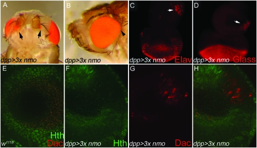
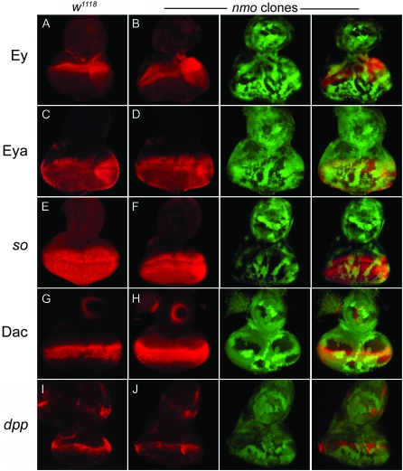
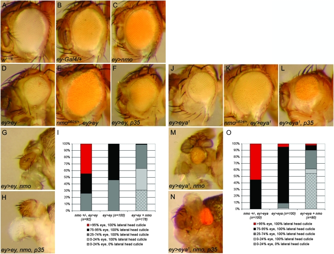
Drosophila nemo (nmo) is the founding member of the Nemo-like kinase (Nlk) family of serine-threonine kinases. Previous work has characterized nmo's role in planar cell polarity during ommatidial patterning. Here we examine an earlier role for nmo in eye formation through interactions with the retinal determination gene network (RDGN). nmo is dynamically expressed in second and third instar eye imaginal discs, suggesting additional roles in patterning of the eyes, ocelli, and antennae. We utilized genetic approaches to investigate Nmo's role in determining eye fate. nmo genetically interacts with the retinal determination factors Eyeless (Ey), Eyes Absent (Eya), and Dachshund (Dac). Loss of nmo rescues ey and eya mutant phenotypes, and heterozygosity for eya modifies the nmo eye phenotype. Reducing nmo also rescues small-eye defects induced by misexpression of ey and eya in early eye development. nmo can potentiate RDGN-mediated eye formation in ectopic eye induction assays. Moreover, elevated Nmo alone can respecify presumptive head cells to an eye fate by inducing ectopic expression of dac and eya. Together, our genetic analyses reveal that nmo promotes normal and ectopic eye development directed by the RDGN.


Figures

F<sc>igure</sc> 1.—
Figure 1.—
The retinal determination gene network. (A) Regulatory interactions within the RDGN. Solid arrows show direct transcriptional regulation, curved arrows demonstrate feedback loops, and dashed lines indicate physical interactions. So, Eya, and Dac are not required for ey expression during normal eye development, but can activate its expression in ectopic eye assays (Pignoni et al. 1997). Modified from Pappu and Mardon (2004) and Silver and Rebay (2005). (B) Schematic of a third instar eye-antennal imaginal disc. The antennal disc gives rise to the antenna and surrounding head cuticle. In the eye disc, the MF marks the dynamic boundary between the posterior, differentiated eye cells and the anterior head primordia. Hh activates dpp transcription in the furrow, which promotes expression of the RD genes and drives the MF forward. Wg, secreted from the anterior dorsal and ventral lobes, promotes head specification by inhibiting furrow progression and transcription of retinal specification genes. Anterior is up; dorsal is left.
F<sc>igure</sc> 2.—
Figure 2.—
nmo is co-expressed with the RDGN in the second instar eye disc. Expression of the nmo-lacZ enhancer trap during second instar eye disc development [63–72 hr after egg laying (AEL)], detected with anti-β-gal antibody. (A) nmo-lacZ is expressed in all cells. (B–G) nmo-lacZ (green, B and E) coincides with Eya (red, C and F) in the posterior eye disc in mid (B–D) and late (E–G) second instar. (H) Schematic summarizing nmo's co-expression with the eye-specification genes. Early: nmo and Ey are co-expressed in the posterior eye field. Mid: nmo is co-expressed with Ey in anterior cells of the eye field and with Ey and Eya in posterior cells. Late: same is in mid, except at the posterior margin where nmo is co-expressed with Ey, Eya, and Dac.
F<sc>igure</sc> 3.—
Figure 3.—
nmo is expressed in multiple cellular contexts in the third instar eye-antennal disc (late third instar:140 hr AEL). All discs are oriented with dorsal left, anterior up. (A–C) nmo-lacZ (green) is coincident with Eya (red) in the MF (arrow) and in the ocellar progenitors (arrowheads) and, to a lesser degree, posterior to the furrow. nmo-lacZ is absent in the PPN domain (bracket). (D–F) nmo-lacZ (green) coincides with Dac (red) in the third antennal disc segment, in addition to the MF and retinal cells. nmo-lacZ overlaps with Dac in the presumptive ocelli (arrowheads in F), although Dac more broadly encompasses the entire dorsal vertex region. (G–I) Hth (red) is absent in eye disc cells expressing nmo-lacZ (red) and reduced in the ocellar primordia (arrowheads in I). (J–L) nmo-lacZ (green) is coincident with Ato (red) in the MF, the ocellar region, and the antennal disc. (M–O) Single confocal section. nmo-lacZ (green) (M) and Hth (red) (N) are expressed in all cells of the PE. (P) Schematic of a third instar eye/antennal disc. The regions of dpp and wg expression and their action on MF progression are shown. The MF moves posterior to anterior. nmo's expression relative to the RD genes and the Wg effector Hth in the eye disc are indicated below, as previously described (Bessa et al. 2002; Silver and Rebay 2005).
F<sc>igure</sc> 4.—
Figure 4.—
nmo modifies the ey small-eye phenotype. (A) Wild-type compound eye. (B) nmoP mutants have narrow eyes and a square ommatidial array. (C) eyR compound eyes are small with disorganized ommatidia and uneven eye margins. The ventral row of sensory vibrissae is often duplicated (arrowhead). The most frequent phenotype is shown. (D) nmoP/+; eyR/+ trans-heterozygotes display a slightly smaller eye compared to wild type. (E) nmoP; eyR/+. The nmoP eye phenotype is not modified by reducing a single copy of eyR. (F) nmoP; eyR. The size and periphery of the compound eye are rescued compared to C. A single set of ventral vibrissae is present (arrowhead), as in wild type. Flies are oriented with the anterior left. The same results were obtained using nmoDB24, nmoadk1, and nmoadk2.
F<sc>igure</sc> 5.—
Figure 5.—
nmo and eya genetically interact. (A) w1118. Sensory vibrissae surround the ventral eye margin (arrowhead). (B). nmoDB24 compound eyes are elongated and narrow. (C) eya2/+; nmoDB24. A secondary eye field develops at the ventral margin (arrow). This phenotype accounts for 22.9% of total observed ventral eye defects (33.6% of flies; n = 104). (D) eya2 mutants lack eyes and are missing ventral vibrissae (arrowhead). As a result of smaller heads, ventral eye bristles converge with dorsal orbital bristles. (E) eya2; nmoDB24/+. More ventral vibrissae are observed (arrowhead; compare with D), and the distance from the dorsal orbital bristles is increased (line). (F) eya2; nmoDB24. The ventral vibrissae have a nearly wild-type pattern (arrowhead), and their distance from the orbital bristles is further rescued from E (line). (G–I) Imaginal eye discs are oriented with the anterior at the top, dorsal left. White images, anti-Cut; red images in H and I, anti-cyclin B. (G) w1118. Anti-Cut labeling (white) marks the antennal disc and anterior-most eye disc cells, which give rise to head cuticle. Additional posterior staining is observed in the PE and ommatidial clusters. (H) eya2. The eye disc is largely reduced, relative to the antennal disc. (I) eya2; nmoDB24. The ventral eye disc is enlarged compared to eya2 (arrowheads near red images), but proliferation (cyclin B labeling, red) is comparable to eya2 alone (H).
F<sc>igure</sc> 6.—
Figure 6.—
nmo potentiates Ey-mediated ectopic eye induction. (A and C) UAS-GFP∷nmo/+; dpp-Gal4/+ (green). (B and D) nmo-lacZ (red). (A and B) dpp-Gal4 (green) targets expression in the dorsal and ventral poles of the eye disc and in a ventral wedge in the antennal eye disc, which bisects nmo-expressing cells (red, B). (C and D) dpp-Gal4 (green) drives expression along the A–P boundary of the wing disc, which intersects with nmo expression (red) at the dorsal wing hinge (boxes). (E) UAS-ey/+; dpp-Gal4/+ wing disc. E′–E″′ is an enlarged view of the dorsal wing pouch in E. Ectopic eyes are induced in cells ectopically expressing Dac (red, E′) and with reduced Hth (green, E″). E″′ is a composite of E′ and E″. (F) nmo-lacZ (blue) wing disc, Dac (red), and Hth (green). F′–F″′ is an enlarged view of the dorsal wing pouch in F. Dac is not normally expressed in the dorsal wing hinge (F′; compare with E′), although Hth (F″) and nmo (F″′) are normally co-expressed in dorsal wing cells able to be respecified to the eye fate (boxes in C–F). (G–J) Eye discs stained for ELAV (red). (G) w1118. ELAV is normally expressed in the posterior photoreceptors and is absent in the antennal disc. (H) UAS-ey/+; dpp-Gal4/+. ELAV-positive ectopic photoreceptors are detected in the ventral antennal disc (arrowhead). Photoreceptors do not differentiate at the dorsal and ventral boundaries of the eye field, where Dpp targets expression (see A), and the size of the eye disc is reduced compared to the antennal disc. (I) UAS-ey/+; nmoDB24/dppGal4. Ectopic photoreceptors are no longer detected in the antennal disc (arrowhead). The normal photoreceptor field, as well as the overall size of the eye/antennal disc, is further reduced compared to H. (J) UAS-ey/UAS-nmo; dpp-Gal4/+. Large groups of ectopic photoreceptors are detected in the ventral antennal disc (arrowhead). The normal eye field is rescued (compare with H). (K and O) UAS-ey/+; dpp-Gal4/+. Ectopic eyes are induced ventrally to the antennae (arrows, K) and on the legs and wing hinge (arrowheads, O). (L and P) UAS-ey/+; dpp-Gal4/nmoDB24. Ectopic eyes are induced at a lower frequency and are smaller than in K (arrow, L). Ectopic eye fields on the legs and wing hinge are reduced compared to O. (M and Q) UAS-ey/+; dpp-Gal4,nmoDB24/nmoDB24. Ectopic eyes are only rarely induced on the head (M). Ectopic eye fields on the leg and wing hinge are considerably reduced (compare with O). The compound eye has the characteristic nmo morphology. (N and R) UAS-ey/UAS-nmo; dpp-Gal4/+. The size of ectopic eyes induced on the head (N) and on the leg and wing hinge (R) are larger than in K and O, respectively. (S) Quantification of the phenotypes in K–N. The relative frequencies of zero, one, or two ectopic eyes on head cuticle derived from the antennal disc for the indicated genotypes. Loss or co-expression of nmo has a dose-dependent effect on both the frequency and the penetrance of the ectopic eye phenotype. Loss of nmo significantly reduces the penetrance of head-to-eye respecification.
F<sc>igure</sc> 7.—
Figure 7.—
nmo potentiates Eya-mediated ectopic eye formation. (A and E) dppGal4/UAS-eya2. (A) Small fields of ectopic eyes are induced on head cuticle below the antennae (arrows). (B and F) nmoDB24, dppGal4/UAS-eya2. (B) Ectopic eye fields are induced less frequently and are smaller than in A (arrow). (C and G) nmoDB24, dppGal4/nmoDB24, UAS-eya2. (C) Ectopic eyes are rarely induced. (D and H). UAS-nmo/+; dppGal4/UAS-eya2. (D) Large ectopic eye fields frequently merge with the endogenous eye (arrows). (E) The compound eye is overgrown (arrowhead). (F) The compound eye has minimal overgrowth (arrowhead; compare with E). (G) The compound eye is smaller than wild type and resembles nmo mutants. (H) The compound eye is massively overgrown (arrowheads). (I) Quantification of phenotypes in A–D. Loss of nmo dose-dependently reduces the frequency of head-to-eye respecification. (J) Quantification of leg-to-eye transformations for the indicated genotypes. As in the head (I), loss of nmo dose-dependently reduces the frequency of ectopic eyes induced on the leg. Co-expression with nmo increases the penetrance and frequency. Flies were reared at 29°.
F<sc>igure</sc> 8.—
Figure 8.—
nmo potentiates Dac-mediated ectopic eye formation. Quantification of the relative frequency of head-to-eye transformations for the genotypes shown. Heterozygosity for nmo reduces the frequency of ectopic eye formation, but less potently than with misexpressed ey (Figure 6S) or eya (Figure 7I).
F<sc>igure</sc> 9.—
Figure 9.—
Ectopic Nmo induces head-to-eye respecification in the antennal disc. (A–D and F–H) UAS-nmo; dppGal4/UAS-nmo (A) Of pharate adults, 16.7% (28/168) display pigmented retinal cells on the antero-ventral head cuticle (arrows). (B) The dorsal eye is overgrown (arrowhead). (C–H). Imaginal eye/antennal discs. (C) Clusters of ELAV-positive cells (arrow) are detected in the antero-ventral head primordia in 63.6% (28/44) of antennal discs. ELAV is not normally expressed in the antennal disc. (D) Eye-antennal discs labeled with anti-Glass, which is normally absent from the antennal disc, indicate that ventral antennal cells have adopted a retinal fate (arrow). (E–H) Confocal images of the mid-ventral antennal disc taken at ×40. E is a Z-stack of the entire antennal disc. F–H are single confocal planes. (E) w1118. Expression of Hth (green) and Dac (red) in a wild-type disc. Hth is ubiquitously expressed in cells of the outer antennal segments and overlaps with Dac (red) in the third antennal segment. (F) Misexpressed Nmo induces loss of Hth (G) and concomitant ectopic Dac (red) in the outer antennal ring. Endogenous Dac expression is below this focal plane. (H) Composite of F and G. (C–H) Imaginal discs are oriented anterior up, dorsal left.
F<sc>igure</sc> 10.—
Figure 10.—
Nmo promotes eye development independently of RDGN gene activation. (A, C, E, G, and I) Wild type. (B, D, F, H, and J) nmoDB24 somatic clones are marked by loss of GFP (green). (C–J) nmo loss-of-function clones have no effect on Ey (A and B), Eya (C and D), so-lacZ (E and F), Dac (G and H), or dpp-lacZ expression (I and J). Eye discs are oriented with the anterior up, dorsal left.
F<sc>igure</sc> 11.—
Figure 11.—
nmo promotes early eye defects associated with ectopic ey and eya. (A) w1118. (B) ey-Gal4/+ and (C) ey-Gal4/UAS-nmo have no detectable external abnormal phenotype. (D) ey-Gal4/UAS-ey causes a smaller, rough eye. (E) ey-Gal4/UAS-ey; nmoDB24/+. Loss of a single copy of nmoDB24 rescues the small eye induced by UAS-ey (compare with D). (F) ey-Gal4/UAS-ey; UAS-p354/+. Blocking apoptosis does not phenocopy loss of nmo (compare with E). (G) eyGal4/UAS-ey; UAS-nmo/+. Flies frequently lack all eye and most head structures. (H) eyGal4/UAS-ey; UAS-nmo/UAS-p35. Blocking apoptosis does not modify the defects induced by UAS-ey and UAS-nmo (compare with G). (I) Quantification of the phenotypes observed in misexpression analysis with UAS-ey (D–H). (J) ey-Gal4/UAS-eya1 results in smaller eyes with dorsal overproliferation. (K) ey-Gal4/UAS-eya1; nmoDB24/+. Loss of a single copy of nmoDB24 rescues the small eye and dorsal overproliferation induced by eya (compare with J). (L) ey-Gal4/UAS-eya1; UAS-p35/+. Blocking apoptosis does not phenocopy loss of nmo (compare with K). (M) ey-Gal4/UAS-eya1; UAS-nmo/+. Flies display severe reduction of the compound eye and head cuticle. (M) ey-Gal4/UAS-eya1; UAS-nmo/UAS-p35. Blocking apoptosis does not modify the defects induced by UAS-eya1 and UAS-nmo (compare with M).(O) Quantification of phenotypes observed in misexpression analysis with UAS-eya1 (J–N).

Similar articles

Cited by

References

    1. Abu-Shaar, M., H. D. Ryoo and R. S. Mann, 1999. Control of the nuclear localization of Extradenticle by competing nuclear import and export signals. Genes Dev. 13 935–945. - PMC - PubMed
    1. Anderson, J., C. L. Salzer and J. P. Kumar, 2006. Regulation of the retinal determination gene dachshund in the embryonic head and developing eye of Drosophila. Dev. Biol. 297 536–549. - PMC - PubMed
    1. Azpiazu, N., and G. Morata, 2000. Function and regulation of homothorax in the wing imaginal disc of Drosophila. Development 127 2685–2693. - PubMed
    1. Azpiazu, N., and G. Morata, 2002. Distinct functions of homothorax in leg development in Drosophila. Mech. Dev. 119 55–67. - PubMed
    1. Baonza, A., and M. Freeman, 2002. Control of Drosophila eye specification by Wingless signalling. Development 129 5313–5322. - PubMed

Publication types

Substances

LinkOut - more resources