. 2008 Oct;10(10):1172-80.
doi: 10.1038/ncb1777. Epub 2008 Aug 31.

Dynein is required for polarized dendritic transport and uniform microtubule orientation in axons

Affiliations

Dynein is required for polarized dendritic transport and uniform microtubule orientation in axons

Yi Zheng et al. Nat Cell Biol. 2008 Oct.

Abstract

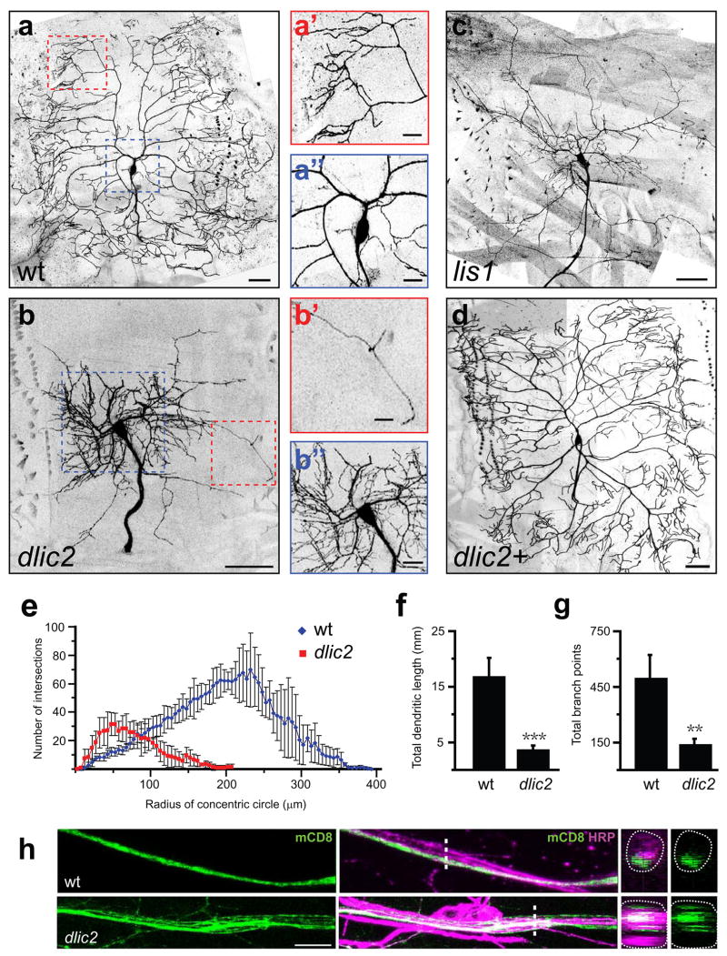
Axons and dendrites differ in both microtubule organization and in the organelles and proteins they contain. Here we show that the microtubule motor dynein has a crucial role in polarized transport and in controlling the orientation of axonal microtubules in Drosophila melanogaster dendritic arborization (da) neurons. Changes in organelle distribution within the dendritic arbors of dynein mutant neurons correlate with a proximal shift in dendritic branch position. Dynein is also necessary for the dendrite-specific localization of Golgi outposts and the ion channel Pickpocket. Axonal microtubules are normally oriented uniformly plus-end-distal; however, without dynein, axons contain both plus- and minus-end distal microtubules. These data suggest that dynein is required for the distinguishing properties of the axon and dendrites: without dynein, dendritic organelles and proteins enter the axon and the axonal microtubules are no longer uniform in polarity.


Figures

Figure 1
Figure 1. dlic21157 Functions Cell-Autonomously to Regulate Dendrite and Axon Development
Dendritic arbours are labelled by mCD8-GFP (anterior is to the left and dorsal is up). Scale bars: 30 μm (a–d) and 10 μm (a′,a″,b′,b″,h). Error bars represent Standard Deviation (S.D.) in this and all subsequent figures. (a–d) Clones of class IV ddaC neurons: (a) wt, (b) dlic21157, (c) lis1G10.14 and (d) dlic21157; dlic2-eGFP (dlic2+ rescue). The size and pattern of the dlic21157 and lis1G10.14 arbours are grossly abnormal (b,c). The number of terminal branches in dlic21157 is substantially reduced (compare a′ to b′) whereas the number of proximal branches is greatly increased (compare a″ to b″). Expressing dlic2-eGFP specifically in dlic21157 clones fully rescues the dendrite and axon phenotypes (d). (e) Sholl analysis of dendritic arbours of wt and dlic21157 ddaC clones. Concentric circles with 5 μm increments were drawn around the soma and the number of dendritic branches that intersected each circle was tallied. In control ddaC neurons the number of branches increases progressively from proximal to distal and the maximal number of dendrite branches are found between 225 and 250 μm from the cell body. The dlic21157 neurons exhibited a dramatic proximal shift in dendrite distribution such that the majority of dendrites are located within 100 μm of the soma. Blue diamonds: wt, red squares: dlic21157. (f) Dendrite length of wt ddaC (16,902 ± 3,292 μm, n=4) and dlic21157 ddaC (3,671 ± 681 μm, n=4, ***p<0.001, Student’s unpaired t- test). The dendrite length of dlic21157 ddaC is greatly reduced. (g) The number of dendritic branch points is decreased in dlic21157 ddaC (140 ± 29, n=4, **p<0.01, Student’s unpaired t- test) compared to wt (499 ± 124, n=4). (h) Axons of individual wt (top) and dlic21157 (bottom) ddaC clones coursing through the intersegmental nerve (ISN). The wt axon is a single process whereas the dlic21157 axon has separated into multiple neurites, as revealed in cross sections of the ISN (rightmost panels) at the points indicated by the dashed line. Dotted lines in the rightmost panels delineate the boundary of the nerve. Green: mCD8; magenta: HRP, which labels all nerve processes.
Figure 2
Figure 2. Loss of Dynein Function Alters Golgi Outpost Distribution
Green: ManII-eGFP, magenta: mCD8. Arrowheads: axon, arrows: position of Golgi outposts. (a–h) Localization of Golgi outposts in wt and dlic21157 ddaC clones. Boxes with dotted lines highlight axons (shown in c,d) and boxes with dashed lines highlight proximal dendrites (shown in e,f). Distal dendritic tips are shown (g,h). Scale bars: 30 μm (a,b) and 15 μm (c–h). (a–f) In (c) wt ddaC there are virtually no Golgi outposts in the axon (box with dotted lines in a). In contrast, many are present in (d) dlic21157 axons (box with dotted lines in b). Golgi outposts are more numerous in the proximal dendritic arbour of dlic21157 clones (compare e and f). The signal intensity is optimized for visualizing the smaller Golgi outposts in the proximal arbour; consequently, the outposts close to the soma are over-exposed (e,f). (g,h) Along the distal dendrite there are fewer Golgi outposts in (h) dlic21157 compared to (g) wt. (i,j) Frames from movies (Supplementary Information, Movies 3 and 4) following Golgi outposts in the soma and axon of (i) control and (j) dic1229/dicts neurons. Time (in seconds) is as indicated. The single Golgi outpost (arrowhead) in the control axon does not move whereas in the dic1229/dicts neuron there are many Golgi outposts in the axon and one Golgi outpost (arrow) moves from the soma into the axon. Scale bar: 12.5 μm. (k) Number of Golgi outposts in proximal (control: 10.00 ± 3.39, n=5; dlic21157: 15.45 ± 4.18, n=11, *p<0.025, Student’s unpaired t- test) and distal (control: 7.92 ± 3.11 per 100 μm, n=6; dlic21157: 2.00 ± 2.00 per 100 μm, n=3, *p<0.025, Student’s unpaired t-test) dendritic arbours as well as axons (control: 2.33 ± 2.71 per 100 μm, n=6; dlic21157: 34.49 ± 14.55 per 100 μm, n=5, ***p<0.001, Student’s unpaired t- test). (l) Size of Golgi outposts in proximal (control: 3.31 ± 1.08 μm2, n=5; dlic21157: 6.00 ± 1.75 μm2, n=11, **p<0.01, Student’s unpaired t- test) and distal (control: 1.12 ± 0.36 μm2, n=6; dlic21157: 0.25 ± 0.25 μm2, n=3; **p<0.01, Student’s unpaired t- test) dendritic arbours as well as axons (control: 0.45 ± 0.51, n=6; dlic21157: 16.71 ± 6.09, n=5, ***p<0.001, Student’s unpaired t- test).
Figure 3
Figure 3. Localization of Endosomes and the Ion Channel Ppk Depends on Dynein
Green: RFP (a–d) or Ppk (e,f), magenta: mCD8. ppk-Gal4 UAS-mCD8-GFP (a–f) was used to visualize dendrites and axons of ddaC neurons expressing the endosomal marker UAS-Rab4-RFP (a,b) or UAS-Spin-RFP (c,d). Arrows indicate dendrites, arrowheads point to axons. Scale bar: 30 μm. (a,b) Rab4-RFP is present in dendrites and axons in (a) wt; however, its dendritic localization is reduced in (b) dic1229/dicts. (c,d) Spinster-RFP localizes to dendrites and axons in (c) wt, but its dendritic distribution is reduced in (d) dic1229/dicts. (e,f) Ppk is found specifically in dendrites in (e) wt, but is present in both axons and dendrites of (f) dlic21157 ddaC clones. (g) Table summarizing the axonal and dendritic localization of Golgi outposts, endosomal markers and Ppk in wt (dynein +) and dynein loss-of-function (dynein-) neurons.
Figure 4
Figure 4. Mislocalization of Nod-βgal, but not Kin-βgal, in Dynein Mutant Neurons
Green: βgal, magenta: mCD8. UAS-nod-lacZ (a–d) and UAS-kin-lacZ (e–h) driven by ppk-Gal4, which also drives UAS-mCD8-GFP expression. Open arrowhead: proximal axon, filled arrowhead: axon shaft; arrows: proximal dendrites. Scale bar: 30 μm. (a–d) Localization of Nod-βgal in (a) wt, (b) dic1229/dicts, (c) dlic21157 ddaC clone and (d) ddaC over-expressing dmn (dmn OE). The lower panels (a′–d′) show the Nod-βgal channel at a slightly higher magnification. In wt axons Nod-βgal enters only the very proximal axon but is not present in the axon shaft. In dic1229/dicts, dlic21157 and dmn OE neurons Nod-βgal extends into the axon shaft. The dic1229/dicts axon shown in (b,b′) is not unusually wide, yet it has strong Nod-βgal signal. (e–h) Localization of Kin-βgal in (e) wt, (f) dic1229/dicts, (g) dlic21157 ddaC clone and (h) dmn OE. Inserts show Kin-βgal channel alone. Kin-βgal is normally localized specifically to axons and this distribution is not changed by a reduction in dynein activity.
Figure 5
Figure 5. Mixed Orientation of Axonal MTs in dic1229/dicts Neurons Revealed by EB1-GFP
Kymographs (a,c) and movie frames (b,d) showing the trajectory of EB1-GFP comets in the axons of ddaC neurons. UAS-EB1-GFP was expressed in class IV neurons by 4-77-Gal4 (a–e) and in class I neurons by 2-21-Gal4 (e). Time (in seconds) is as indicated. The bar to the right of the kymogaph indicates the portion of the movie from which the frames were taken. The soma is to the left. (a,c) Kymographs showing that EB1-GFP always moves away from the soma in (a) control axons, but moves both towards and away from the soma in the axons of (c) dic1229/dicts neurons. A portion of the dic1229/dicts movie was out of focus (109–225 sec) and this section of the kymograph was removed for clarity. (b,d) Single frames from movies showing the movement of individual EB1-GFP comets. Arrowheads and diamonds indicate comets moving anterogradely (yellow) or retrogradely (magenta). Scale bar: 5 μm. In (b) control axons all EB1-GFP comets move anterogradely whereas in (d) dic1229/dicts axons EB1-GFP moves both anterogradely and retrogradely. EB1-GFP comets moved in both directions in dic1229/dicts axons with or without ectopic neurites, and within individual neurites EB1-GFP comets moved retrogradely (42%) and anterogradely (58%). (e) Bar graph illustrating the percentage of EB1-GFP comets that move anterogradely (yellow) or retrogradely (magenta) in the axons of class IV and class I control and dic1229/dicts neurons in 3rd instar larvae.

Comment in

  • Dynein branches out.
    Tear G. Tear G. Nat Cell Biol. 2008 Oct;10(10):1131-2. doi: 10.1038/ncb1008-1131. Nat Cell Biol. 2008. PMID: 18830220

References

    1. Hirokawa N, Takemura R. Molecular motors and mechanisms of directional transport in neurons. Nat Rev Neurosci. 2005;6:201–214. - PubMed
    1. Baas PW, Deitch JS, Black MM, Banker GA. Polarity orientation of microtubules in hippocampal neurons: uniformity in the axon and nonuniformity in the dendrite. Proc Natl Acad Sci U S A. 1988;85:8335–8339. - PMC - PubMed
    1. Baas PW, Vidya Nadar C, Myers KA. Axonal transport of microtubules: the long and short of it. Traffic. 2006;7:490–498. - PubMed
    1. Sharp DJ, et al. Identification of a microtubule-associated motor protein essential for dendritic differentiation. J Cell Biol. 1997;138:833–843. - PMC - PubMed
    1. Yu W, et al. Depletion of a microtubule-associated motor protein induces the loss of dendritic identity. J Neurosci. 2000;20:5782–5791. - PMC - PubMed

Publication types