. 2008 Oct;9(10):1157-64.
doi: 10.1038/ni.1645. Epub 2008 Aug 31.

Toll-like receptor-mediated induction of type I interferon in plasmacytoid dendritic cells requires the rapamycin-sensitive PI(3)K-mTOR-p70S6K pathway

Affiliations

Toll-like receptor-mediated induction of type I interferon in plasmacytoid dendritic cells requires the rapamycin-sensitive PI(3)K-mTOR-p70S6K pathway

Weiping Cao et al. Nat Immunol. 2008 Oct.

Abstract

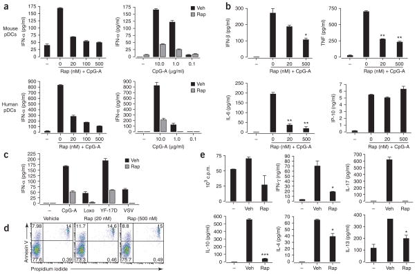
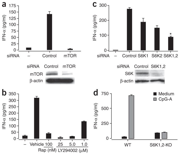
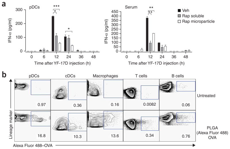
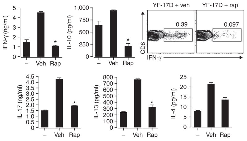
Robust production of type I interferon (IFN-alpha/beta) in plasmacytoid dendritic cells (pDCs) is crucial for antiviral immunity. Here we show involvement of the mammalian target of rapamycin (mTOR) pathway in regulating interferon production by pDCs. Inhibition of mTOR or its 'downstream' mediators, the p70 ribosomal S6 protein kinases p70S6K1 and p70S6K2, during pDC activation by Toll-like receptor 9 (TLR9) blocked the interaction of TLR9 with the adaptor MyD88 and subsequent activation of the interferon-regulatory factor IRF7, which resulted in impaired IFN-alpha/beta production. Microarray analysis confirmed that inhibition of mTOR by the immunosuppressive drug rapamycin suppressed antiviral and anti-inflammatory gene expression. Consistent with this, targeting rapamycin-encapsulated microparticles to antigen-presenting cells in vivo resulted in less IFN-alpha/beta production in response to CpG DNA or the yellow fever vaccine virus strain 17D. Thus, mTOR signaling is crucial in TLR-mediated IFN-alpha/beta responses by pDCs.


Figures

Figure 1
Figure 1
Rapamycin inhibits TLR-mediated IFN-α secretion by pDCs. (a) ELISA of IFN-α in supernatants of isolated pDCs pretreated for 3 h with various doses of rapamycin (Rap) and then stimulated with CpG-A (10 μg/ml; left) or treated with vehicle (Veh) or rapamycin and then stimulated with various doses of CpG-A (right). (b) ELISA of IFN-β, TNF, IL-6 and CXCL10 in supernatants of purified mouse pDCs pretreated with various doses of rapamycin and then stimulated for 24 h with CpG-A (10 μg/ml). (c) ELISA of IFN-α in supernatants of purified spleen pDCs cultured for 24 h with CpG-A (10 μg/ml), loxoribine (Loxo; 1 mM), YF-17D or vesicular stomatitis virus (VSV; all viruses at a multiplicity of infection of 1) after pretreatment with rapamycin (Rap) or vehicle. (d) Apoptotic death of isolated mouse pDCs pretreated with vehicle or 20 nM or 500 nM rapamycin, assessed by staining with annexin V and propidium iodide. Numbers in quadrants indicate percent cells in each. (e) Proliferation of and cytokine induction in naive CD4+CD62L+ OT-II T cells (1 × 10⁵ cells/ well) cultured with pDCs (5 × 10⁴) pretreated with ovalbumin peptide (10 μg/ml) and CpG-A (10 μg/ml) plus vehicle or rapamycin (100 nM for 3 h), assessed after 3 d by incorporation of [3H]thymidine (top left; proliferation) and ELISA of IFN-γ, IL-4, IL-10, IL-13 and IL-17. *, P < 0.05; **, P < 0.001; ***, P < 0.0001. Data are representative of at least three independent experiments (error bars, s.d.).
Figure 2
Figure 2
Expression of rapamycin-sensitive mTOR pathway components in pDCs. (a) Immunoblot analysis of phosphorylated (phospho-) and total mTOR in lysates of RAW cells (4 × 10⁶) transiently transfected for 40 h with cyan fluorescent protein–tagged MyD88 and IRF7-YFP, treated with vehicle (−) or rapamycin (+) and then stimulated for 0–60 min with CpG-A–DOTAP. Results are representative of two independent experiments. (b) Immunoblot analysis of phosphorylated and total mTOR, p70S6K, 4E-BP1 and Akt in lysates of purified pDCs (1 × 10⁶) stimulated for 15 min with CpG-A in the presence (Rap) or absence (Medium) of rapamycin. Results are representative of three independent experiments.
Figure 3
Figure 3
TLR-mediated induction of IFN-α in pDCs depends on the mTOR signaling pathway. (a) ELISA of IFN-α in supernatants (above) and immunoblot analysis of mTOR in lysates (below) of purified mouse pDCs (5 × 10⁵) transfected for 5 h with control or mTOR-specific siRNA and then stimulated for 24 h with CpG-A. Below, β-actin serves as a loading control. (b) ELISA of IFN-α in supernatants of pDCs treated with rapamycin or the PI(3)K inhibitor LY294002 (1–25 μM), then stimulated with CpG-A and assessed 24 h later. (c) ELISA of IFN-α in supernatants (above) and immunoblot analysis of p70S6K in lysates (below) of mouse pDCs transfected for 5 h with siRNA pools specific for p70S6K1 (S6K1), p70S6K2 (S6K2) or both (S6K1,2), then stimulated for 24 h with CpG-A. *, P < 0.05. (d) ELISA of IFN-α in supernatants of pDCs isolated from wild-type (WT) and Rps6k1−/−Rps6k2−/− double-knockout (S6K1,2-KO) mouse spleens and stimulated in vitro with CpG-A. Data are representative of three independent experiments (a–c) or two independent experiments with at least two mice per group per experiment (d); error bars, s.e.m. of replicate wells.
Figure 4
Figure 4
Rapamycin does not affect TLR9 expression or interaction with CpG-A. (a) Flow cytometry of isolated spleen pDCs stimulated with CpG-A with or without rapamycin pretreatment, then stained for intracellular TLR9. (b) Deconvolution microscopy of RAW cells expressing YFP-tagged TLR9 (green), pretreated with vehicle (left) or rapamycin (right) and then incubated for 90 min with indodicarbocyanine-tagged CpG-A–DOTAP (red; 5 μg/ml). Two images were obtained for each condition. Data are representative of two independent experiments.
Figure 5
Figure 5
Rapamycin inhibits the spatial interaction of TLR9 with the MyD88-IRF7 complex and affects the phosphorylation and nuclear translocation of IRF7. (a) Immunoassay of RAW cells (4 × 10⁶) transfected with hemagglutinin-tagged MyD88 and YFP-tagged TLR9 and then, 36 h later, stimulated for 90 min with CpG-A–DOTAP; cell extracts were immunoprecipitated (IP) with anti-hemagglutinin and analyzed by immunoblot (IB) for coprecipitated TLR9. MyD88 serves as a loading control. (b) Flow cytometry of human pDCs pretreated with rapamycin (left) or transfected with control (con) siRNA or siRNA specific for p70S6K1 and p70S6K2 (S6K), then stimulated for 45 min with CpG-A; cells were fixed, made permeable and stained with phycoerythrin-conjugated mouse antibody to IRF7 phosphorylated at Ser477 and Ser479 (phosphor-IRF7) or control isotype antibody (Control Ab; phycoerythrin-conjugated mouse immunoglobulin G1). (c) Confocal microscopy of purified mouse pDCs stimulated for 12 h with CpG-A in the presence of rapamycin or vehicle, then fixed with formaldehyde, made permeable with saponin, blocked with 20% (vol/vol) goat serum, stained with anti-IRF7 (green) and mounted with ProLong Gold antifade reagent with DAPI (blue). Scale bar, 2 μm. (d) Luciferase assay of HEK293 cells transiently transfected with 0 ng (−), 40 ng or 200 ng of constitutively active IRF7 (IRF7-D477,479) together with luciferase-tagged IFN-β (plasmid p125-Luc; 50 ng), renilla luciferase (0.5 ng), wild-type IRF7 (3 ng), MyD88 (20 ng) and TLR9 (50 ng), cultured for 24 h, then pretreated for 3 h with 100 nM rapamycin and then stimulated overnight with CpG-A (10 μg/ml). Empty vector, vector without IRF7. Data are representative of three (a,d) or two (b,c) independent experiments (error bars (d), s.d.).
Figure 6
Figure 6
In vivo administration of rapamycin inhibits YF-17D-induced production of IFN-α in the serum. (a) ELISA of IFN-α production by pDCs from C57BL/6 mice treated daily for 3 d with soluble rapamycin (1.5 mg per kg body weight per day) or rapamycin encapsulated in PLGA microparticles (2 mg per mouse per day), then vaccinated subcutaneously with YF-17D on day 4; blood and spleens were obtained at various times after injection for analysis of serum (right) and pDCs enriched from spleens with microbeads coated with anti–mouse PDCA1 (left). *P < 0.05; **P < 0.001; ***P < 0.0001. Data are representative of at least three independent experiments (error bars, s.d.). (b) Flow cytometry of ovalbumin (OVA) in cells from C57BL/6 mice left untreated (top row) or subcutaneously injected with Alexa Fluor 488–labeled ovalbumin encapsulated in PLGA microparticles (bottom row); draining lymph nodes were isolated 24 h later and treated for 30 min at 37 °C with collagenase type IV, then isolated cells were stained for cell surface lineage markers to identify cell type (above plots). Numbers below outlined areas indicate percent cells containing OVA. cDCs, conventional DCs. Data are representative of three independent experiments.
Figure 7
Figure 7
Inhibition of mTOR in APCs in vivo can suppress the adaptive immune response to a vaccine. IFN-γ production (top right) and cytokine expression (top left and bottom) by CD8+ T cells enriched from draining lymph nodes of C57BL/6 mice left untreated (−) or treated daily for 3 d with vehicle or rapamycin microparticles (2 mg per mouse per day) and then vaccinated subcutaneously on day 4 with YF-17D; 5 d later, single-cell suspensions were cultured for an additional 3 d in vitro in medium alone (left bars) or with a CD8+ T cell epitope from YF-17D (middle and right bars). IFN-γ was assessed by intracellular cytokine staining and flow cytometry; numbers above outlined areas indicate percent CD8+IFN-γ+ cells. IFN-γ, IL-4, IL-13, IL-10 and IL-17 in cell supernatants were measured by ELISA. *, P < 0.05. Data are representative of three independent experiments (error bars, s.d.).

References

    1. Liu YJ. IPC: professional type 1 interferon-producing cells and plasmacytoid dendritic cell precursors. Annu Rev Immunol. 2005;23:275–306. - PubMed
    1. Colonna M, Trinchieri G, Liu YJ. Plasmacytoid dendritic cells in immunity. Nat Immunol. 2004;5:1219–1226. - PubMed
    1. Asselin-Paturel C, et al. Mouse type I IFN-producing cells are immature APCs with plasmacytoid morphology. Nat Immunol. 2001;2:1144–1150. - PubMed
    1. Kawai T, Akira S. Innate immune recognition of viral infection. Nat Immunol. 2006;7:131–137. - PubMed
    1. Ito T, Wang YH, Liu YJ. Plasmacytoid dendritic cell precursors/type I interferon-producing cells sense viral infection by Toll-like receptor (TLR) 7 and TLR9. Springer Semin Immunopathol. 2005;26:221–229. - PubMed

Publication types

MeSH terms