. 2008 Aug 28;59(4):608-20.
doi: 10.1016/j.neuron.2008.06.026.

A positive feedback signal transduction loop determines timing of cerebellar long-term depression

Affiliations

A positive feedback signal transduction loop determines timing of cerebellar long-term depression

Keiko Tanaka et al. Neuron. .

Abstract

Synaptic activity produces short-lived second messengers that ultimately yield a long-term depression (LTD) of cerebellar Purkinje cells. Here, we test the hypothesis that these brief second messenger signals are translated into long-lasting biochemical signals by a positive feedback loop that includes protein kinase C (PKC) and mitogen-activated protein kinase. Histochemical "epistasis" experiments demonstrate the reciprocal activation of these kinases, and physiological experiments--including the use of a light-activated protein kinase--demonstrate that such reciprocal activation is required for LTD. Timed application of enzyme inhibitors reveals that this positive feedback loop causes PKC to be active for more than 20 min, allowing sufficient time for LTD expression. Such regenerative mechanisms may sustain other long-lasting forms of synaptic plasticity and could be a general mechanism for prolonging signal transduction networks.


Figures
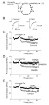
Figure 1
Figure 1. Ca2+ is upstream of PKC, MAPK, and PLA2
(A) Positive feedback loop model for LTD. AA, arachidonic acid; other abbreviations in text. (B) Superimposed PF-EPSCs recorded before and 20 min after uncaging Ca2+ in the absence (left) or presence (right) of intracellular PKCI (10 µM). (C) Time course of LTD induced by uncaging Ca2+ (open circles, n = 7). LTD was blocked by including PKCI in the intracellular solution (closed circles, n = 6). PF-EPSC amplitudes are normalized to their mean pre-uncaging level. (D and E) Ca2+-triggered LTD also was blocked by the MAPK inhibitors PD98059 (100 µM; closed circles in (D), n = 5) and U0126 (20 µM; closed triangles in (D), n = 4), as well as the PLA2 inhibitor OBAA (5 µM; closed circles in (E), n = 5). DMSO, the solvent for these drugs, did not affect LTD (open circles in (D) and (E), n = 7). Error bars in this and subsequent figures indicate ± 1SEM.
Figure 2
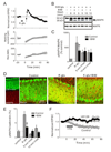
Figure 2. MAPK is activated during LTD and is required downstream of PKC
(A) Time course of LTD induced by K-glu treatment (upper panel, n = 4). Holding current (middle panel) and membrane resistance (lower panel) were changed upon K-glu treatment, but were gradually recovered. (B) Immunoblots detecting phosphorylated MAPK (arrow head in upper panel) and total MAPK (lower panel) in cerebellar slices. Bands at 50 kD in upper panel are immunoglobulin. (C) Stimulation of slices with K-glu led to a sustained increase in phosphorylated MAPK (pMAPK) that was reduced by the PKC inhibitor, BIM (0.2 µM; n = 5–8). (D) Images of cerebellar slices double-stained with antibodies against pMAPK (red) and calbindin (green). (E) Quantification of pMAPK labeling in Purkinje cells (15 fields from 3 experiments). (F) LTD induced by 0.5 µM TPA (open circles, n = 6) was blocked by 20 µM U0126 (closed circles, n = 5). Asterisks indicate significant difference (p < 0.05) between control and BIM treatment.
Figure 3
Figure 3. PKC is activated during LTD induction
(A) Images of Purkinje cell soma in cerebellar slices double-stained with antibodies against PKCα (red) and calbindin (green). (B) Line-scan profiles of calbindin (upper) and PKCα (lower) staining in Purkinje cell bodies. Intensity of PKCα staining at the membrane (m) and center of cell body (c) was measured. (C) PKC translocation, quantified as T = (m−c)/(m+c) × s100 (27–67 cells from 6 experiments). Asterisks indicate significant difference (p < 0.05) between control and U0126 (20 µM) treatment.
Figure 4
Figure 4. MAPK is activated by uncaging MEK-CA
(A) Phosphorylation of purified MAPK protein by caged or uncaged MEK-CA (3 ng/µl) in vitro. (B) Concentration-dependence of MAPK phosphorylation (pMAPK) by caged and uncaged MEK-CA (n = 5). (C) MEK-KD (10 ng/µl) was unable to phosphorylate MAPK protein in vitro even after uncaging, while uncaged MEK-CA (10 ng/µl) strongly phosphorylated MAPK protein.
Figure 5
Figure 5. MAPK activation by uncaging MEK-CA is capable of inducing LTD, which requires PKC
(A) Measurement of EPSC amplitude during dialysis of caged MEK-CA into a Purkinje cell (n = 18). At time 0, whole-cell patch clamp recording was established with an intracellular solution including caged MEK-CA. EPSC amplitude was normalized by dividing by the mean amplitude of EPSCs measured in each cell during the first 10 min of recording. (B) LTD is induced by uncaging MEK-CA (n = 23) but not by uncaging MEK-KD (n = 4). (C) Induction of LTD by uncaging MEK-CA prevents further depression by pairing PF stimulation with Purkinje cell depolarization (PF&ΔV, n = 4). (D) The induction of LTD by PF&ΔV also prevents further depression by uncaging MEK-CA (n = 3). (E) Summarized data of averaged reduction of PF-EPSC in occlusion experiments. Once LTD was induced by uncaging MEK-CA or PF&ΔV (open columns), subsequent LTD induction (closed columns) produced no further depression of PF-EPSCs. Asterisks indicate significant difference (p < 0.05). (F) The LTD induced by uncaging MEK-CA is blocked by 10 µM PKCI (red, n = 6) or 5 µM OBAA (blue, n = 6). Control data are same as in (B).
Figure 6
Figure 6. Sustained activation of PKC is required to induce LTD
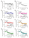
(A) BIM treatment (0.5 µM) had no effect on the amplitude of PF-EPSCs (n = 4). (B) LTD induced by PF&ΔV in control conditions (n = 9). (C–H) The effect of external application of BIM at various times after PF&ΔV (black bars) on LTD (closed circles; n = 3–6). Control responses shown in (B), recorded in the absence of BIM, are overlaid for comparison in each panel (open circles).
Figure 7
Figure 7. Sustained activation of PLA2 is required to induce LTD
(A) OBAA treatment (5 µM) had no effects on the amplitude of PF-EPSCs (n = 7). (B) LTD induced by PF&ΔV in control conditions (n = 6). (C–F) LTD measured after applying OBAA in the external solution at the indicated times (closed circles; n = 4–5) after PF&ΔV (horizontal bars). Control LTD shown in (B) is overlaid in each panel (open circles).
Figure 8
Figure 8. Sustained activation of PKC and PLA2 is required for the LTD
(A and B) Mean LTD, measured 45–55 min after stimulation, are plotted as a function of the time when BIM (A) or OBAA (B) were applied. Data are normalized to LTD measured in control conditions and lines indicate linear regression fits.
Figure 9
Figure 9
Model for time course of signal transduction events involved in LTD. LTD is initiated (blue) when synaptic activity generates short-lived second messengers such as Ca2+, which leads to activation of the positive feedback loop (red) that causes induction of LTD by phosphorylating AMPAR and leading to changes in AMPAR trafficking. At late times, synthesis of unknown new proteins leads to long-term maintenance of LTD (green).

References

    1. Ahn S, Ginty DD, Linden DJ. A late phase of cerebellar long-term depression requires activation of CaMKIV and CREB. Neuron. 1999;23:559–568. - PubMed
    1. Alessi DR, Cuenda A, Cohen P, Dudley DT, Saltiel AR. PD 098059 is a specific inhibitor of the activation of mitogen-activated protein kinase kinase in vitro and in vivo. J. Biol. Chem. 1995;270:27489–27494. - PubMed
    1. Anderson WW, Collingridge GL. The LTP program: a data acquisition program for on-line analysis of long-term potentiation and other synaptic events. J. Neurosci. Methods. 2001;108:71–83. - PubMed
    1. Artola A, Singer W. Long-term depression of excitatory synaptic transmission and its relationship to long-term potentiation. Trends Neurosci. 1993;16:480–487. - PubMed
    1. Atkins CM, Davare MA, Oh MC, Derkach V, Soderling TR. Bidirectional regulation of cytoplasmic polyadenylation element-binding protein phosphorylation by Ca2+/calmodulin-dependent protein kinase II and protein phosphatase 1 during hippocampal long-term potentiation. J. Neurosci. 2005;25:5604–5610. - PMC - PubMed

Publication types

MeSH terms

Substances

LinkOut - more resources