. 2008 Sep;83(3):408-14.
doi: 10.1016/j.ajhg.2008.08.003. Epub 2008 Sep 4.

C-terminal deletions in the ALAS2 gene lead to gain of function and cause X-linked dominant protoporphyria without anemia or iron overload

Affiliations

C-terminal deletions in the ALAS2 gene lead to gain of function and cause X-linked dominant protoporphyria without anemia or iron overload

Sharon D Whatley et al. Am J Hum Genet. 2008 Sep.

Abstract

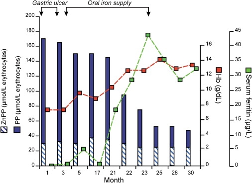
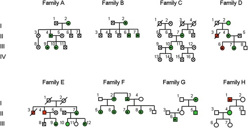
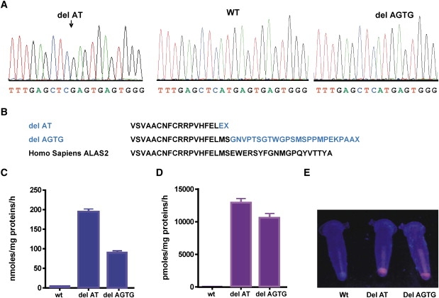
All reported mutations in ALAS2, which encodes the rate-regulating enzyme of erythroid heme biosynthesis, cause X-linked sideroblastic anemia. We describe eight families with ALAS2 deletions, either c.1706-1709 delAGTG (p.E569GfsX24) or c.1699-1700 delAT (p.M567EfsX2), resulting in frameshifts that lead to replacement or deletion of the 19-20 C-terminal residues of the enzyme. Prokaryotic expression studies show that both mutations markedly increase ALAS2 activity. These gain-of-function mutations cause a previously unrecognized form of porphyria, X-linked dominant protoporphyria, characterized biochemically by a high proportion of zinc-protoporphyrin in erythrocytes, in which a mismatch between protoporphyrin production and the heme requirement of differentiating erythroid cells leads to overproduction of protoporphyrin in amounts sufficient to cause photosensitivity and liver disease.


Figures

Figure 1
Figure 1
Treating Iron Deficiency Decreases Erythrocyte Protoporphyrin Concentrations Iron deficiency caused by bleeding from a gastric ulcer was treated with omeprazole and oral iron.
Figure 2
Figure 2
Pedigrees of Eight Families with FECH-Mutation-Negative Protoporphyria Green circles and squares represent patients with photosensitivity. Red circles and squares represent patients with photosensitivity and clinically overt liver disease. Protoporphyric liver disease was confirmed at autopsy or by needle biopsy in all these patients except patient I1 (family H), for whom a diagnosis has not been established. Patient II3 (family E) has been reported previously. Clinical information was not obtainable for patients C I, 3 and 4 or E I, 1 and 2. Black dots within circles or squares indicate individuals in whom either the delAGTG (families A–E, H) or delAT (families F and G) ALAS2 mutations were identified. Crosses within circles or squares indicate individuals in whom sequencing excluded the presence of an ALAS2 mutation. The absence of a black dot or cross indicates an individual from whom a DNA sample was not available for analysis. The LOD score for linkage between photosensitivity and the ALAS2 mutation was calculated for families A–E.
Figure 3
Figure 3
C-Terminal Deletions in ALAS2 Cause X-Linked Dominant Protoporphyria (A) Sequence analysis of genomic DNA from male patients showing deletions in the ALAS2 gene. (B) Predicted effects of deletions on ALAS2 C-terminal sequences. (C–E) Prokaryotic expression of wild-type and mutant ALAS2 enzymes: Rates of formation of ALA (C) and porphyrin (D) by bacterial lysates; means and ranges for three experiments are shown. (E) Porphyrin fluorescence (UVA light) in bacterial pellets.
Figure 4
Figure 4
Comparison of C-terminal Sequences of ALAS enzymes (A) Alignment of ALAS C-terminal sequences. (B) Phylogenetic tree showing relationships between ALAS genes constructed with ClustalW.

References

    1. Sassa S. Modern diagnosis and management of the porphyrias. Br. J. Haematol. 2006;135:281–292. - PubMed
    1. Bottomley S.S. Sideroblastic anemias. In: Lukens J.N., Rogers G.M., Paraskevas F., Glader B.E., editors. Wintrobe's Clinical Hematology, J.P. Greer J. Foerster. Lippincott Williams & Wilkins; Philadelphia: 2004. pp. 1012–1033.
    1. Cox T.M. Erythropoietic protoporphyria. In: Kadish K.M., Smith K.M., Guilard R., editors. The Porphyrin Handbook, Vol 14, Medical aspects of porphyrias. Academic Press; Amsterdam: 2003. pp. 121–150.
    1. Gouya L., Martin-Schmitt C., Robreau A.M., Austerlitz F., Da Silva V., Brun P., Simonin S., Lyoumi S., Grandchamp B., Beaumont C. Contribution of a common single-nucleotide polymorphism to the genetic predisposition for erythropoietic protoporphyria. Am. J. Hum. Genet. 2006;78:2–14. - PMC - PubMed
    1. Whatley S.D., Mason N.G., Holme S.A., Anstey A.V., Elder G.H., Badminton M.N. Gene dosage analysis identifies large deletions of the FECH gene in 10% of UK families with erythropoietic protoporphyria. J. Invest. Dermatol. 2007;127:2790–2794. - PubMed

Publication types