. 2009 Oct;19(4):565-72.
doi: 10.1111/j.1750-3639.2008.00201.x. Epub 2008 Aug 29.

Hippocampal sclerosis: progress since Sommer

Affiliations

Hippocampal sclerosis: progress since Sommer

Maria Thom. Brain Pathol. 2009 Oct.

Abstract

Hippocampal sclerosis (HS) continues to be the most common pathology identified in patients with refractory temporal lobe epilepsy undergoing surgery. Wilhelm Sommer described this characteristic pattern of neuronal loss over 120 years ago through his post-mortem studies on patients with epilepsy. Neuropathological post-mortem studies in the 20th century proceeded to contribute significantly to the understanding of this disease process, with regard to the varying patterns of HS and involvement of adjacent limbic structures. From studies of surgical temporal lobe specimens from the 1950s onwards it was recognized that an early cerebral injury could act as the precipitant for the sclerosis and epilepsy. Modern neuropathological studies have focused on aspects of neuronal injury, loss of specific neuronal groups and cellular reorganization to address mechanisms of epileptogenesis and the enigma of how specific hippocampal neuronal vulnerabilities and glial proliferation are both the effect and the cause of seizures.


Figures

Figure 1
Figure 1
Sommer's description of the vulnerable region of neuronal loss in Ammon's horn sclerosis that became known as Sommer's sector. In his original paper he describes this as “The whole part of the grey matter that belongs to the medial wall of the lower horn, approximately from the fold‐over locus of the ventricular endothelium over the middle of the curvature . . . or if one considers the Ammon's horn as an ellipsoid, whose biggest axis goes parallel to the horizontal plane, in a sector in which the lower exterior quadrant (calculated from the medial plane of the brain) covers approximately 60 degrees of the horizontal axis towards the smaller one, and 20 degrees in the upper exterior.” In the figure these parameters have been superimposed on a surgical resection specimen immunostained with neuronal nuclear antigen to highlight Sommer's description.
Figure 2
Figure 2
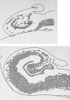
One of the first illustrations of hippocampal sclerosis from an original paper published in 1987 by Bratz (8) depicting the regions of neuronal loss and reduction in volume in the hippocampus compared with a control. Note that the granule cell layer also appears somewhat broader than in the control although the phenomena of granule cell dispersion in the dentate gyrus remained to be described until 1990.
Figure 3
Figure 3
Illustrations taken from the 2nd edition of Greenfield (33) in the chapter on epilepsy authored by Alfred Meyer showing (A) severe degeneration in the amygdala (arrows), (B) Ammon's horn sclerosis involving Sommer's sector and (C) temporal neocortex with severe laminar neuronal loss from mainly cortical layer three (indicated by arrows). This pattern of temporal lobe neocortical sclerosis, with loss of neurons from the superficial cortical layers, is more easily visible in lobectomy specimens with neuronal nuclear antigen immunohistochemistry, as illustrated in (D). The reproduction of figures from Greenfield's Neuropathology, 2nd Edn., is with permission from Hodder Education.
Figure 4
Figure 4
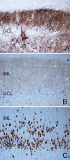
Mossy fiber sprouting, as described by Sutula in 1989, can be seen in the molecular layer of the dentate gyrus in hippocampal sclerosis using (A) Timms method or (B) dynorphin immunohistochemistry. Dispersion of the granule cells of the dentate gyrus in hippocampal sclerosis is also a common finding and was first described by Houser in 1990 and is illustrated here with neuronal nuclear antigen staining (C). Abbreviations: GCL = granule cell layer; IML = inner molecular layer.

Similar articles

Cited by

References

    1. Aronica E, Gorter JA (2007) Gene expression profile in temporal lobe epilepsy. Neuroscientist 13:100–108. - PubMed
    1. Aronica E, Gorter JA, Ramkema M, Redeker S, Ozbas‐Gerceker F, Van Vliet EA et al (2004) Expression and cellular distribution of multidrug resistance‐related proteins in the hippocampus of patients with mesial temporal lobe epilepsy. Epilepsia 45:441–451. - PubMed
    1. Aronica E, Boer K, Van Vliet EA, Redeker S, Baayen JC, Spliet WG et al (2007) Complement activation in experimental and human temporal lobe epilepsy. Neurobiol Dis 26:497–511. - PubMed
    1. Babb TL, Brown WJ, Pretorius J, Davenport C, Lieb JP, Crandall PH (1984) Temporal lobe volumetric cell densities in temporal lobe epilepsy. Epilepsia 25:729–740. - PubMed
    1. Blumcke I, Thom M, Wiestler OD (2002) Ammon's horn sclerosis: a maldevelopmental disorder associated with temporal lobe epilepsy. Brain Pathol 12:199–211. - PMC - PubMed

Personal name as subject