

The relationship between inflammation and new bone formation in patients with ankylosing spondylitis
- PMID: 18761747
- PMCID: PMC2592781
- DOI: 10.1186/ar2496
The relationship between inflammation and new bone formation in patients with ankylosing spondylitis
Abstract
Introduction: Spinal inflammation as detected by magnetic resonance imaging and new bone formation as identified by conventional radiographs are characteristic of ankylosing spondylitis. Whether and how spondylitis and syndesmophyte formation are linked are unclear. Our objective was to investigate whether and how spinal inflammation are associated with new bone formation in ankylosing spondylitis.
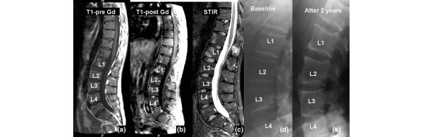
Methods: Spinal magnetic resonance images and conventional radiographs from 39 ankylosing spondylitis patients treated with anti-tumour necrosis factor (anti-TNF) agents at baseline and after 2 years were analysed for syndesmophyte formation at vertebral edges with or without inflammatory lesions at baseline.
Results: Overall, 922 vertebral edges at the cervical and lumbar spine were analysed. At baseline, the proportion of vertebral edges with and without inflammation (magnetic resonance imaging) that showed structural changes (conventional radiographs) was similar (in total, 16.6% of all vertebral edges in 71.4% of patients). From the perspective of syndesmophyte formation (n = 26, 2.9%) after 2 years, there were more vertebral edges without (62%) than with (38%) inflammation at baseline (P = 0.03). From the perspective of spinal inflammation at baseline (n = 153 vertebral edges), more syndesmophytes developed at vertebral edges with (6.5%) than without (2.1%) inflammation (P = 0.002, odds ratio 3.3, 95% confidence interval 1.5 to 7.4). Inflammation persisted in 31% of the initially inflamed vertebral edges (n = 132), and new lesions developed in 8% of the vertebral edges without inflammation at baseline (n = 410). From the perspective of spinal inflammation after 2 years (n = 72 vertebral edges), 5.6% of the vertebral edges showed syndesmophyte development in contrast to 1.9% of the vertebral edges with new syndesmophytes without inflammation (P = 0.06).
Conclusions: These findings obtained in patients treated with anti-TNF agents suggest linkage and some dissociation of inflammation and new bone formation in ankylosing spondylitis. Although syndesmophytes were also found to develop at sites where no inflammation had been seen by magnetic resonance imaging at baseline, it was more likely that syndesmophytes developed in inflamed vertebral edges. More effective suppression of spinal inflammation may be required to inhibit structural damage in ankylosing spondylitis.
Figures
References
-
- Baraliakos X, Landewe R, Hermann KG, Listing J, Golder W, Brandt J, Rudwaleit M, Bollow M, Sieper J, Heijde D van der, Braun J. Inflammation in ankylosing spondylitis: a systematic description of the extent and frequency of acute spinal changes using magnetic resonance imaging. Ann Rheum Dis. 2005;64:730–734. doi: 10.1136/ard.2004.029298. - DOI - PMC - PubMed
-
- Wanders A, Landewe R, Dougados M, Mielants H, Linden S van der, Heijde D van der. Association between radiographic damage of the spine and spinal mobility for individual patients with ankylosing spondylitis: can assessment of spinal mobility be a proxy for radiographic evaluation? Ann Rheum Dis. 2005;64:988–994. doi: 10.1136/ard.2004.029728. - DOI - PMC - PubMed
MeSH terms
Substances
LinkOut - more resources
Full Text Sources
Medical
Research Materials
