. 2008;10(5):R105.
doi: 10.1186/ar2497. Epub 2008 Sep 1.

Elevated autoantibody content in rheumatoid arthritis synovia with lymphoid aggregates and the effect of rituximab

Affiliations

Elevated autoantibody content in rheumatoid arthritis synovia with lymphoid aggregates and the effect of rituximab

Sanna Rosengren et al. Arthritis Res Ther. 2008.

Abstract

Introduction: The purpose of this study was to quantitatively evaluate the contribution of synovial lymphoid aggregates to autoantibody (rheumatoid factor [RF] and anti-cyclic citrullinated peptide [anti-CCP]) and total immunoglobulin (IgG and IgM) production in rheumatoid arthritis (RA) patients and the effect thereon of the B-cell-depleting antibody, rituximab, in the ARISE (Assessment of Rituximab's Immunomodulatory Synovial Effects) trial.

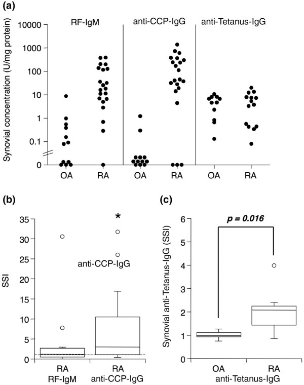
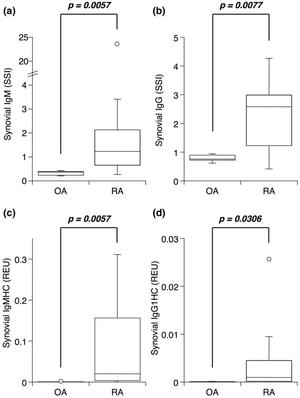
Methods: Autoantibodies as well as total IgM and IgG were quantified by enzyme-linked immunosorbent assay in extracts of synovial tissues and matched serum from patients with RA or osteoarthritis (OA). Synovial biopsies and serum were obtained at baseline and 8 weeks following rituximab therapy in 14 RA patients. A synovial/serum index (SSI) was calculated as the ratio of synovial to serum antibody/albumin, with values above 1 representing synovial enrichment. Lymphoid aggregates were evaluated histologically.

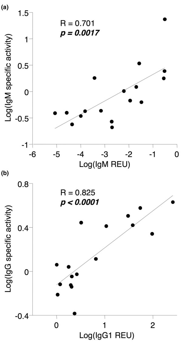
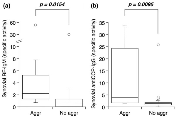
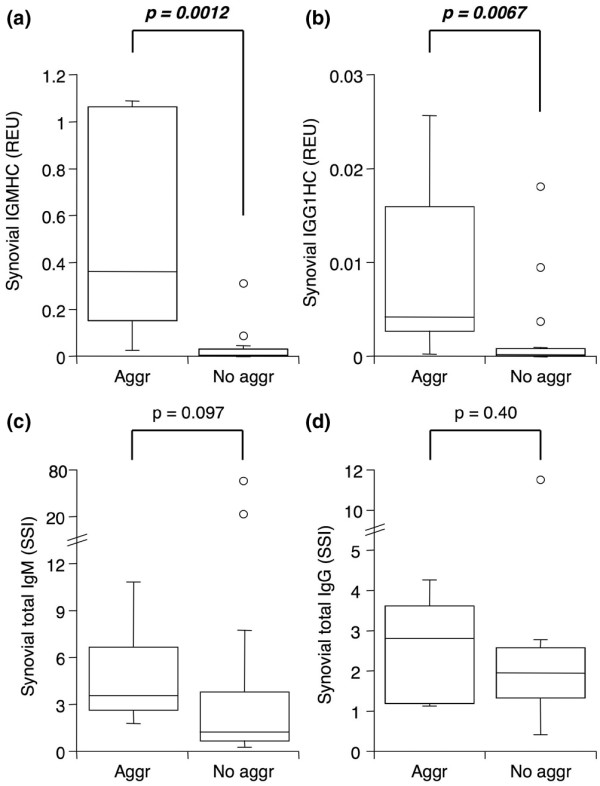
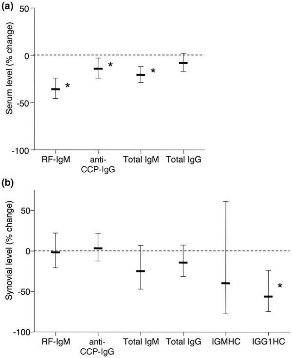
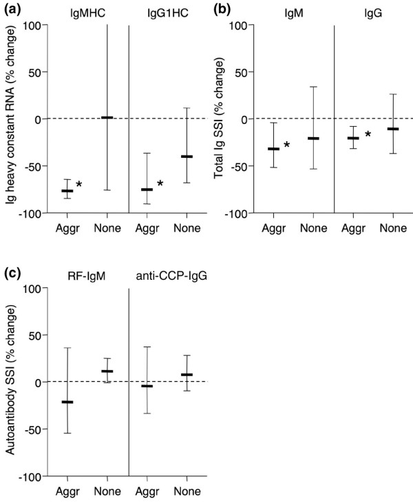
Results: Anti-CCP IgG, but not RF-IgM, was significantly enriched in RA synovia compared with serum. Total IgM and IgG were also enriched in RA, but not in OA. SSI correlated significantly with mRNA content for both IgM and IgG, demonstrating that it reflected synovial immunoglobulin production. RA synovia with lymphocyte aggregates contained significantly elevated RF-IgM and anti-CCP IgG compared with tissues with diffuse lymphoid infiltration. Rituximab treatment did not affect synovial autoantibody or total immunoglobulin SSI overall. However, in aggregate-containing tissues, rituximab significantly reduced total IgM and IgG SSI as well as IgM and IgG1 mRNA. Surprisingly, RF-IgM and anti-CCP IgG SSIs were unchanged by rituximab in aggregate-containing synovia.

Conclusions: Combined with earlier observations that synovial lymphoid aggregates are unaltered by rituximab treatment, these data suggest that lymphoid aggregates may provide a protective niche for autoantibody-producing cells.

Trial registration: ClinicalTrials.gov NCT00147966.


Figures

Figure 1
Figure 1
Detection and enrichment of autoantibodies in rheumatoid arthritis (RA) synovial extracts. Tissues were obtained by arthroplasty or arthroscopic shaver biopsy, and serum and synovial extracts were analyzed by enzyme-linked immunosorbent assay for antibodies of interest and albumin. (a) Individual levels of RF-IgM, anti-CCP IgG, and anti-tetanus IgG in extracts from osteoarthritis (OA) (n = 12, autoantibodies; n = 11, anti-tetanus) and RA (n = 21, autoantibodies; n = 14, anti-tetanus) synovial tissue, normalized to total protein concentration. Limits of detection are 4 (RF-IgM), 10 (anti-CCP IgG), and 0.3 (anti-tetanus IgG). Serum-normalized levels of (b) RF-IgM and anti-CCP IgG in RA synovial extracts (n = 11) and (c) anti-tetanus IgG in OA (n = 6) and RA (n = 9) synovial extracts. In the box (interquartile range, IQR) and whisker (maximum and minimum) plots, the horizontal line inside the box denotes median and the unfilled circles denote outliers outside IQR ± 1.5 × IQR. The asterisk denotes P = 0.019 by Wilcoxon sign rank test to 1 (no enrichment) for anti-CCP IgG (a), and the indicated P value was determined by Wilcoxon rank sum test between OA and RA for anti-tetanus IgG (b). The value for RF-IgM was not significantly above 1 (P = 0.32). anti-CCP, anti-cyclic citrullinated peptide; RF-IgM, rheumatoid factor of the IgM subtype; SSI, synovial/serum index.
Figure 2
Figure 2
Significant enrichment of total IgM and IgG in rheumatoid arthritis (RA) synovia. Serum-normalized levels of (a) total IgM and (b) total IgG in extracts from osteoarthritis (OA) (n = 6) and RA (n = 11) synovial tissue obtained by arthroplasty or arthroscopic shaver biopsy and measured by enzyme-linked immunosorbent assay for antibodies of interest and albumin. GAPDH-normalized message for IgM (c) and IgG1 (d) heavy constant region as determined by quantitative real-time polymerase chain reaction in the same synovia as in (a) and (b). See Figure 1 legend for box plot definitions. Indicated P values were determined by Wilcoxon rank sum test. GAPDH, glyceraldehyde-3-phosphate dehydrogenase; REU, relative expression units; SSI, synovial/serum index.
Figure 3
Figure 3
Correlation between synovial/serum index (SSI) and mRNA for total IgM and IgG. Correlation plots for SSI versus GAPDH-normalized mRNA levels for total IgM (a) and total IgG (b) in rheumatoid arthritis (RA) and osteoarthritis (OA) synovia. Data are from Figure 2. Correlation coefficients (R) and P values were determined by Spearman rank correlation. GAPDH, glyceraldehyde-3-phosphate dehydrogenase; REU, relative expression units.
Figure 4
Figure 4
The presence of lymphoid aggregates in rheumatoid arthritis (RA) synovia is associated with elevated synovial autoantibody levels. Serum-normalized levels of (a) RF-IgM and (b) anti-CCP IgG in extracts from RA synovia with (Aggr, n = 8) or without (No aggr, n = 17) lymphoid aggregates. Tissues were obtained by arthroplasty or arthroscopic shaver biopsy, and extracts were analyzed by enzyme-linked immunosorbent assay for antibodies of interest and albumin. See Figure 1 legend for box plot definitions. Indicated P values were determined by Wilcoxon rank sum test. anti-CCP, anti-cyclic citrullinated peptide; RF-IgM, rheumatoid factor of the IgM subtype.
Figure 5
Figure 5
The presence of lymphoid aggregates in rheumatoid arthritis (RA) synovia is associated with elevated total immunoglobulin message, but not protein. GAPDH-normalized message for IgM (a) and IgG1 (b) heavy constant region in cDNA from RA synovia with (Aggr, n = 8) or without (No aggr, n = 17) lymphoid aggregates. Serum-normalized levels of (c) total IgM and (d) total IgG in extracts from the same synovia as in (a) and (b). Tissues were obtained by arthroplasty or arthroscopic shaver biopsy, and cDNA was analyzed by quantitative real-time polymerase chain reaction (a, b). Extracts were analyzed by enzyme-linked immunosorbent assay for antibodies of interest and albumin (c, d). See Figure 1 legend for box plot definitions. Indicated P values were determined by Wilcoxon rank sum test. GAPDH, glyceraldehyde-3-phosphate dehydrogenase; REU, relative expression units; SSI, synovial/serum index.
Figure 6
Figure 6
Effect of rituximab on circulating and synovial autoantibodies and immunoglobulin. (a) Circulating levels of autoantibodies and total IgM and IgG 8 weeks after rituximab treatment are expressed as percentage of pretreatment levels. (b) Serum-normalized levels of autoantibodies and total IgM and IgG determined by enzyme-linked immunosorbent assay, or mRNA levels of IgM and IgG1 heavy constant region determined by quantitative real-time polymerase chain reaction, in synovial biopsies 8 weeks after rituximab treatment are expressed as percentage of pretreatment levels. Data are expressed as geometric mean ± 95% confidence interval (CI) of 14 subjects. Asterisks denote that 95% CI excludes 0% change (stippled line). anti-CCP, anti-cyclic citrullinated peptide; RF-IgM, rheumatoid factor of the IgM subtype.
Figure 7
Figure 7
Rituximab selectively lowers total immunoglobulin synthesis but not autoantibody content in rheumatoid arthritis (RA) synovia containing lymphoid aggregates. (a) GAPDH-normalized synovial message for IgM and IgG1 heavy constant region in RA synovial biopsies with (Aggr, n = 5) or without (None, n = 9) lymphoid aggregates 8 weeks after rituximab treatment. Serum-normalized levels of (b) total IgM and total IgG and (c) RF-IgM and anti-CCP IgG in extracts from the same synovia as in (a). Data are expressed as geometric mean ± 95% confidence interval (CI) of post-treatment levels relative to pretreatment levels. Asterisks denote that 95% CI excludes 0% change (stippled line). anti-CCP, anti-cyclic citrullinated peptide; GAPDH, glyceraldehyde-3-phosphate dehydrogenase; RF-IgM, rheumatoid factor of the IgM subtype; SSI, synovial/serum index.

Similar articles

Cited by

References

    1. Rantapaa-Dahlqvist S, de Jong BA, Berglin E, Hallmans G, Wadell G, Stenlund H, Sundin U, van Venrooij WJ. Antibodies against cyclic citrullinated peptide and IgA rheumatoid factor predict the development of rheumatoid arthritis. Arthritis Rheum. 2003;48:2741–2749. doi: 10.1002/art.11223. - DOI - PubMed
    1. Klareskog L, Stolt P, Lundberg K, Kallberg H, Bengtsson C, Grunewald J, Ronnelid J, Harris HE, Ulfgren AK, Rantapaa-Dahlqvist S, Eklund A, Padyukov L, Alfredsson L. A new model for an etiology of rheumatoid arthritis: smoking may trigger HLA-DR (shared epitope)-restricted immune reactions to autoantigens modified by citrullination. Arthritis Rheum. 2006;54:38–46. doi: 10.1002/art.21575. - DOI - PubMed
    1. Edwards JC, Szczepanski L, Szechinski J, Filipowicz-Sosnowska A, Emery P, Close DR, Stevens RM, Shaw T. Efficacy of B-cell-targeted therapy with rituximab in patients with rheumatoid arthritis. N Engl J Med. 2004;350:2572–2581. doi: 10.1056/NEJMoa032534. - DOI - PubMed
    1. Leandro MJ, Edwards JC, Cambridge G. Clinical outcome in 22 patients with rheumatoid arthritis treated with B lymphocyte depletion. Ann Rheum Dis. 2002;61:883–888. doi: 10.1136/ard.61.10.883. - DOI - PMC - PubMed
    1. Cambridge G, Leandro MJ, Edwards JC, Ehrenstein MR, Salden M, Bodman-Smith M, Webster AD. Serologic changes following B lymphocyte depletion therapy for rheumatoid arthritis. Arthritis Rheum. 2003;48:2146–2154. doi: 10.1002/art.11181. - DOI - PubMed

Publication types

MeSH terms

Associated data