. 2008 Oct 23;26(45):5741-51.
doi: 10.1016/j.vaccine.2008.08.020. Epub 2008 Aug 30.

Local and humoral immune responses against primary and repeat Neisseria gonorrhoeae genital tract infections of 17beta-estradiol-treated mice

Affiliations

Local and humoral immune responses against primary and repeat Neisseria gonorrhoeae genital tract infections of 17beta-estradiol-treated mice

Wenxia Song et al. Vaccine. .

Abstract

The 17beta-estradiol-treated mouse model is the only small animal model of gonococcal genital tract infection. Here we show gonococci localized within vaginal and cervical tissue, including the lamina propria, and high numbers of neutrophils and macrophages in genital tissue from infected mice. Infection did not induce a substantial or sustained increase in total or gonococcal-specific antibodies. Mice could be reinfected with the same strain and repeat infection did not boost the antibody response. However, intravaginal immunization of estradiol-treated mice induced gonococcal-specific primary and secondary serum antibody responses. We conclude that similar to human infection, experimental murine infection induces local inflammation but not an acquired immune response or immunological memory.


Figures

Fig. 1
Fig. 1
Colonization of the murine lower genital tract by N. gonorrhoeae. Two days after estradiol pellet implantation and antibiotic treatment, mice were inoculated intravaginally with 1 × 10⁶CFU of N. gonorrhoeae strain FA1090 (infected) or the same volume of PBS (uninfected). Genital tissues were collected at days 2 and 5 post-inoculation, embedded in paraffin after fixation in 10% neutral buffered formalin. Tissue sections (5–7 µm) were probed with mAb (H5), which is specific against gonococcal porin followed by HRP-conjugated secondary antibody, and visualized with HRP substrate 3,3-diaminobenzidine in the presence of hydrogen peroxide, followed by hematoxylin and eosin staining. Shown are represented images of H&E staining only (A and B) and antibody plus H&E staining (C and D). Sections were analyzed under a light microscope using a 100× oil objective and numbers of antibody-positive gonococci in five randomly selected fields were estimated. The data are plotted as the average number of bacteria among 100 cells from five mice of each group (E).
Fig. 2
Fig. 2
Increased PMNs in the genital tissues of infected mice. Paraffin embedded sections from tissues of (A and B) infected mice and (C and D) uninfected controls were stained with hematoxylin and eosin. Images were acquired using 100× oil objectives.
Fig. 3
Fig. 3
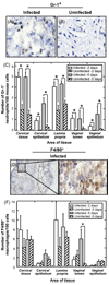
Recruitment of neutrophils and macrophages to genital tissues. Cryosections from tissues of infected and uninfected control mice were stained with (A and B) Gr-1- or (D and E) F4/80-specific antibody and HRP-conjugated secondary antibodies. The sections were further stained with hematoxylin. Images were acquired using 100× oil objective. Control experiments where the primary antibody was omitted did not show significant staining.(C and F)The number of cells with positive staining on five randomly selected microscopic fields of each area in each mouse was counted. The data are plotted as the average number of positively stained cells among 100 mouse cells (±S.D.) based on five mice in each group. *P< 0.05.
Fig. 4
Fig. 4
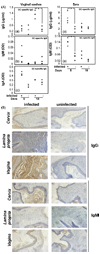
Humoral responses against gonococcal lower genital tract infection. (A) Vaginal washes and sera were collected from infected and control mice on days 5 and 10 post-inoculation. Protease inhibitors were added to prevent protein degradation. The concentration of gonococcal-specific IgG, IgM, and IgA were determined by ELISA.For gonococcal-specific IgG, purified mouse Mab H5 of known concentrations was used to establish a standard curve. Open circles represent data for individual mice and the averages are represented by horizontal bars. *P< 0.05. (B) Paraffin-embedded sections from tissues of infected and uninfected control mice were stained with HRP-conjugated goat-anti-mouse IgG or IgM antibody. The sections were further stained with hematoxylin. Images were acquired using 10× objectives. Control experiments using irrelevant goat antibodies that were conjugated to HRP did not show significant staining.
Fig. 5
Fig. 5
Serum estradiol levels in mice treated with a high dose, slow-release pellet versus a lower total dose of estradiolws. Groups of mice were either given (A) three 0.5 mg injections of estradiolws spaced two days apart or (B) a 5 mg, 21 days slow-release estradiol pellet. Serum estradiol levels were determined at the time points indicated. Mice were not challenged with N. gonorrhoeae. The day on which mice normally would be inoculated with gonococci is two days after estradiol pellet implantation or 6 h after the second dose of estradiolws. Normal serum estradiol levels range from 10 to 130 pg/ml over the course of the estrous cycle [21].
Fig. 6
Fig. 6
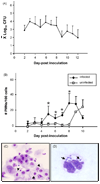
Kinetics of colonization and PMN response in mice treated with 17β-estradiolws. BALB/c mice were treated with 17β-estradiolws and antibiotics and inoculated intravaginally with 1 × 10⁶ CFU N. gonorrhoeae FA1090 (infected), or PBS (control) as described in the Materials and methods. (A) Recovery of N. gonorrhoeae over time expressed as log10 CFU within a 100 µl of vaginal swab suspension [±standard error (S.E.)]. (B) Average number of PMNs out of 100 vaginal cells in stained smears from test mice (diamonds) and mice inoculated with saline (squares) (±S.E.). Representative images of stained vaginal smears from infected mice show (C) epithelial cell-associated gonococci on day 3 post-bacterial challenge and (D) gonococci within a PMN on day 5 of infection. The lesser impact of using 17β-estradiolws on vaginal histology is reflected by a predominance of nucleated epithelial cells as opposed to mostly squamous epithelial cells in panel C.
Fig. 7
Fig. 7
Susceptibility of mice to repeat gonococcal infection. Test mice were inoculated with N. gonorrhoeae strain FA1090 (1 × 10⁶ CFU) using the estradiolws protocol (round one, primary infection). Ten days after infection, mice were treated with ceftriaxone. Four weeks later mice were re-treated with 17β-estradiolws and challenged with 107 CFU of the same strain of N. gonorrhoeae (round two, repeat infection). Age-matched, estradiol-treated mice were challenged with PBS (round one) followed by N. gonorrhoeae (primary infection, round two) in parallel to test mice, and served as naïve controls. Vaginal mucus was quantitatively cultured each day after bacterial challenge. (A) The duration of infection (last positive culture day) from individual test mice (rounds 1 and 2) and naïve control mice (round 2), and represents the combined results of two experiments, each of which were similar. The mean duration of infection is indicated by the horizontal bars with the actual value indicated in parenthesis. (B)The average number (±S.E.) of bacteria in vaginal specimens from five mice in one of the two experiments shown in panel A. (C) Vaginal washes and sera were collected from test and naïve control mice on days 5 and 10 of the second round of infection. Protease inhibitors were added to prevent protein degradation. The concentration of gonococcal-specific IgG were determined by ELISA. Purified mouse Mab H5 of known concentrations was used to establish a standard curve.

Similar articles

Cited by

References

    1. Gerbase AC, Rowley JT, Heymann DH, Berkley SF, Piot P. Global prevalence and incidence estimates of selected curable STDs. Sex Transm Infect. 1998;74 June (Suppl. 1):S12–S16. - PubMed
    1. McNabb SJ, Jajosky RA, Hall-Baker PA, Adams DA, Sharp P, Worshams C, et al. Summary of notifiable diseases—United States, 2006. MMWR Morb Mortal Wkly Rep. 2008;55(March (53)):1–92. - PubMed
    1. Hook EW, Handsfield HH. Gonococcal Infections in the adult. In: Holmes KKMP, Sparling PF, Lemon SM, Stamm WE, Piot P, Wasserheit JN, editors. Sexually transmitted diseases. New York: McGraw-Hill; 1999. pp. 451–466.
    1. Tapsall JW. Antibiotic resistance in Neisseria gonorrhoeae. Clin Infect Dis. 2005;41 August (Suppl. 4):S263–S268. - PubMed
    1. Cohen MS, Hoffman IF, Royce RA, Kazembe P, Dyer JR, Daly CC, et al. Reduction of concentration of HIV-1 in semen after treatment of urethritis: implications for prevention of sexual transmission of HIV-1. AIDSCAP Malawi Research Group. Lancet. 1997;349(June (9069)):1868–1873. - PubMed

Publication types

MeSH terms